Virus Replication

Summary

Many enveloped viruses are released from infected cells by maturing and budding at the plasma membrane. During this process, components of the viral core are incorporated into membrane vesicles that contain viral transmembrane proteins, called “spike” proteins. For many years, these spike proteins, which are required for infectivity, were believed to be incorporated into virions through direct interaction between their cytoplasmic domains and components of the viral core.

More recent evidence shows that while such direct interactions drive budding in alphaviruses, this may not be the case for negative-stranded RNA viruses and retroviruses. These viruses can generate particles in the absence of spike proteins, using only viral core components to drive the process. In some cases, the spike proteins, without the viral core, can be released as virus-like particles. Therefore, optimal sprouting and release may depend on a concerted ‘push and pull action of the core and spike, where oligomerization of both components plays a crucial role.

As viruses are obligate intracellular pathogens, they cannot replicate without the machinery and metabolism of a host cell. Although the replicative life cycle of viruses differs greatly between virus species and categories, there are six basic stages that are essential for viral replication.

1. Adhesion: Viral proteins on the capsid or phospholipid envelope interact with specific receptors on the host cell surface. This specificity determines the host range (tropism) of a virus.

2. Penetration: The process of binding to a specific receptor can induce conformational changes in the viral capsid proteins, or the lipid envelope, resulting in fusion of the viral and cellular membranes. Some DNA viruses can also enter the host cell through receptor-mediated endocytosis.

3. Stripping: The viral capsid is removed and degraded by viral enzymes or host enzymes that release the viral genomic nucleic acid.

4. Replication: After the viral genome has been unwrapped, transcription or translation of the viral genome begins. It is this stage of viral replication that differs greatly between DNA and RNA viruses and viruses with opposite nucleic acid polarity. This process culminates in the de novo synthesis of viral proteins and genome.

5. Assembly: After de novo synthesis of the viral genome and proteins, which can be modified post-transcriptionally, the viral proteins are packaged with the newly replicated viral genome into new virions that are ready to be released from the host cell. This process can also be called maturation.

6. Virion release: There are two methods of viral release: lysis or budding. Lysis results in the death of an infected host cell, these types of viruses are called cytolytic. One example is variola major, also known as smallpox. Enveloped viruses, such as the influenza A virus, are usually released from the host cell by budding. It is this process that results in the acquisition of the phospholipid viral envelope. These types of viruses do not usually kill the infected cell and are called cytopathic viruses.

After virion release, some viral proteins remain within the host cell membrane, serving as potential targets for circulating antibodies. Residual viral proteins that remain within the host cell cytoplasm can be processed and presented at the cell surface on MHC class I molecules, where they are recognized by T cells.

Materials And Methods

  • Viruses and cells.

Vero and HeLa cells were maintained in Dulbecco’s Modified Eagle’s Medium (Invitrogen) supplemented with 5% fetal bovine serum, 5% bovine serum, penicillin and streptomycin. Virus stocks were prepared by infecting confluent monolayers of Vero cells with the KOS strain of HSV type 1 or the Becker strain of pseudorabies virus (PRV) at a multiplicity of infection of 0.01 for 1 h.

Why envelope forms in some kind of the viruses only? What is enzyme and mechanism behind it to form an envelope? - Quora

After infection, cells were incubated in Dulbecco’s modified Eagle’s medium supplemented with 2% fetal bovine serum, 25 mM HEPES buffer, glutamine (0.3 μg/ml), penicillin, and streptomycin until a cytopathic effect was visualized. complete (~3 days). Cells were then scraped into the medium, frozen (-80 °C) and thawed (37 °C) three times, and then sonicated (4 °C) at moderate power for three pulses of 1 min. Cellular debris was removed by centrifugation and aliquots were frozen at -80°C until use.

  • Mutant viruses.

The GC-null mutant and revertant (GC-R) used in these studies were propagated in Vero cells as described previously (kindly provided by Harvey Friedman). The GB null mutants (KO82 and F-BAC GB-) and complementary cells (VB38) used for their growth were generously provided by David Johnson. To generate GB-deficient virions, the progeny of GB-depleted viruses were obtained after infection of non-complementary Vero cells. The ΔUL16 mutant used in the supporting studies (see Supplementary Material) was a gift from Joel Baines and has been described previously.

  • MEN treatment of herpesviruses.

Extracellular virions harvested between 18 and 24 h post-infection were treated with 10 mM NEM for 30 min at 37 °C, either before or after unwrapping with NP-40 (final concentration, 0.5 %. ; I3021; ​​Sigma). Previous work revealed that this concentration and treatment time efficiently modify free cysteines, and in our system also maintain the UL16-capsid interaction in virions.

After NEM treatment, virions and capsids were pelleted at 83,500 × g for 1 hour and samples were separated on 10% sodium dodecyl sulfate (SDS) polyacrylamide gels and electroblotted onto nitrocellulose membranes. The enhanced chemiluminescence method of immunoblot analysis was performed according to the manufacturer’s instructions (Amersham). Rabbit anti-HSV UL16 and anti-VP5 sera were used at dilutions of 1:6,000 and 1:7,500, respectively. PRV UL16-specific antibodies were kindly provided by Thomas Mettenleiter and used at a dilution of 1:15,000.

  • Virus-cell binding assay.

Confluent monolayers of cells in 100-mm dishes were incubated with 1 mL of virus stock (∼5 × 10⁸ PFU) at 4°C with rocking for 45 min to allow virus attachment. NEM was added to the virus before or after incubation with cells for 30 min at 4°C. Unbound virus and residual NEM were removed by washing the cells with 5 ml of phosphate-buffered saline (PBS).

Viral membranes that remained attached to the cells were solubilized with 4 mL of NP-40 lysis buffer (0.5% NP-40, 150 mM NaCl, 50 mM Tris-HCl [pH 8.0]) for 15 min. at 37°C.  Lysates were removed from the dish by pipetting, and insoluble material and cell debris were removed by centrifugation for 10 min at 3,829 × g. The supernatant capsids were pelleted through a 700 μl 30% sucrose cushion (wt/vol in TNE [20 mM Tris-HCl {pH 7.6}, 100 mM NaCl, 1 mM EDTA]) in a rotor. SW55ti at 83,500 × g for 1 h.

Pellets were dissolved in SDS sample buffer (2% SDS, 62.5 mM Tris-HCl, 10% glycerol, 0.025% bromophenol blue, 50 mM dithiothreitol [DTT], and 5% β-mercaptoethanol [BME]), boiled and separated by SDS. – polyacrylamide gel electrophoresis (PAGE) in 10% gels, and analyzed by immunoblotting using specific antibodies for VP5 and UL16. The amount of UL16 was determined by densitometry and normalized to VP5 levels.


Prion Diseases

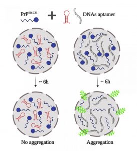
Prion, which is an abnormal form of a normally harmless protein found in the brain that is responsible for a variety of fatal neurodegenerative diseases in animals, including humans, is called transmissible spongiform encephalopathies.

In the early 1980s, American neurologist Stanley B. Prusiner and his colleagues identified the “infectious protein particle,” a name that was shortened to “prion” (pronounced “pree-on”). Prions can enter the brain through infection or can arise from mutations in the gene that codes for the protein.

Once present in the brain, prions multiply by inducing benign proteins to fold into an abnormal shape. This mechanism is not fully understood, but another protein normally found in the body may also be involved. Normal protein structure is thought to consist of a series of flexible coils called alpha-helices. In prion protein, some of these helices are stretched out into flat structures called beta-strands.

The normal conformation of the protein can be degraded quite easily by cellular enzymes called proteases, but the prion protein form is more resistant to this enzymatic activity. Therefore, as prion proteins multiply, they are not broken down by proteases and instead accumulate inside neurons, destroying them. The progressive destruction of neurons eventually causes brain tissue to fill with holes in a sponge-like or spongiform pattern.

Prion diseases that affect humans include: Creutzfeldt-Jakob disease, Gerstmann-Sträussler-Scheinker disease, fatal familial insomnia, and kuru. Prion diseases that affect animals include scrapie, bovine spongiform encephalopathy (commonly called mad cow disease), and chronic wasting of mule deer and elk.

For decades, doctors thought these diseases were the result of infection with slow-acting viruses, named for the long incubation times required for the diseases to develop. These diseases were called, and are sometimes still called slow infections. The pathogen of these diseases has certain viral attributes, such as extremely small size and strain variation, but other properties are atypical of viruses. In particular, the agent is resistant to ultraviolet radiation, which normally inactivates viruses by destroying their nucleic acid.

Prions differ from all other known disease-causing agents in that they appear to lack nucleic acid, that is, DNA or RNA, which is the genetic material that all other organisms contain. Another unusual feature of prions is that they can cause hereditary, infectious and sporadic diseases; for example, Creutzfeldt-Jakob disease manifests itself in all three forms, with sporadic cases being the most common.

Prion proteins can act as infectious agents, spreading disease when transmitted to another organism, or they can arise from an inherited mutation. Prion diseases also show a sporadic pattern of incidence, meaning that they appear to appear randomly in the population. The underlying molecular process that causes the prion protein to form in these cases is unknown. Other neurodegenerative disorders, such as Alzheimer’s disease or Parkinson’s disease, may arise from molecular mechanisms that result in protein misfolding that are similar to those that cause prion diseases.

Types of prion diseases

Prion disease can occur in both humans and animals. Below are some different types of prion diseases.

Human prion diseases

  • Creutzfeldt-Jakob disease (CJD). First described in 1920, CJD can be acquired, inherited, or sporadic. Most CJD cases are sporadic.
  • Variant Creutzfeldt-Jakob disease (vCJD). This form of CJD can be acquired by eating the contaminated meat of a cow.
  • Fatal familial insomnia (FFI). FFI affects the thalamus, which is the part of your brain that manages your sleep-wake cycles. One of the main symptoms of this condition is worsening insomnia. The mutation is dominantly inherited, meaning an affected person has a 50 per cent chance of passing it on to their children.
  • Gerstmann-Straussler-Scheinker syndrome (GSS). GSS is also inherited. Like FFI, it is dominantly transmitted. It affects the cerebellum, which is the part of the brain that handles balance, coordination, and equilibrium.
  • Kuru: Kuru was identified in a group of people from New Guinea. The disease was transmitted through a form of ritual cannibalism in which the remains of deceased relatives were consumed.

Risk factors for these diseases include:

  • Genetics. If someone in your family has an inherited prion disease, you are also at higher risk of having the mutation.
  • Years. Sporadic prion diseases tend to develop in older adults.
  • Animal products. Eating animal products that are contaminated with a prion can give you a prion disease.
  • Medical procedures. Prion diseases can be transmitted through contaminated medical equipment and nerve tissue. Cases, where this has happened, include transmission through contaminated corneal transplants or dura mater grafts.

What are the symptoms of prion disease?

Prion diseases have very long incubation periods, often on the order of many years. When symptoms develop, they get progressively worse, sometimes rapidly. Common symptoms of prion disease include:

  • difficulties with thinking, memory, and judgment
  • personality changes such as apathy, agitation, and depression
  • confusion or disorientation
  • involuntary muscle spasms (myoclonus)
  • loss of coordination (ataxia)
  • difficulty sleeping (insomnia)
  • difficult or slurred speech
  • vision problems or blindness

How is prion disease treated?

There is currently no cure for prion disease. But treatment focuses on providing supportive care. Examples of this type of care include:

  • Medications: Some medications may be prescribed to help treat symptoms. Examples include:

– reduce psychological symptoms with antidepressants or sedatives
– providing pain relief using opiate medications
– relieve muscle spasms with drugs such as sodium valproate and clonazepam

  • Assistance. As the disease progresses, many people need help to care for themselves and do their daily activities.
  • Providing hydration and nutrients. In the advanced stages of the disease, intravenous fluids or a feeding tube may be required.

Malaria

Overview

Malaria is a disease caused by a parasite. The parasite is transmitted to humans through the bites of infected mosquitoes. People who have malaria usually feel very sick with a high fever and chills. Although the disease is rare in temperate climates, malaria remains common in tropical and subtropical countries. Every year, almost 290 million people become infected with malaria and more than 400,000 people die from the disease.

To reduce malaria infections, global health programs distribute preventive medicines and insecticide-treated nets to protect people from mosquito bites. The World Health Organization has recommended a malaria vaccine for use in children living in countries with a high number of malaria cases.

Protective clothing, mosquito nets, and insecticides can protect you while you travel. You can also take preventive medicine before, during, and after a trip to a high-risk area. Many malaria parasites have developed resistance to the common drugs used to treat the disease.

Symptoms

Malaria signs and symptoms may include:

  • Fever
  • Shaking chills
  • General feeling of discomfort.
  • Headache
  • nausea and vomiting
  • Diarrhea
  • Abdominal pain
  • muscle or joint pain
  • Fatigue
  • Fast breathing
  • fast heart rate
  • Cough

Some people who have malaria experience cycles of malaria “attacks.” An attack usually begins with chills and chills, followed by a high fever, sweats, and a return to normal temperature. The signs and symptoms of malaria usually begin a few weeks after being bitten by an infected mosquito. However, some types of malaria parasites can remain dormant in your body for up to a year.

When to see a doctor

Talk to your doctor if you experience a fever while living in or after travelling to a high-risk malaria region. If you have severe symptoms, seek emergency medical attention.

Causes

Malaria is caused by a unicellular parasite of the genus Plasmodium. The parasite is most commonly transmitted to humans through mosquito bites.

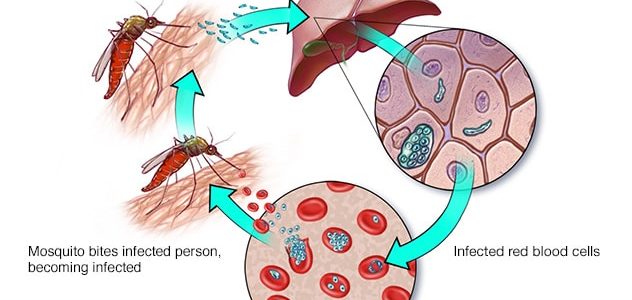
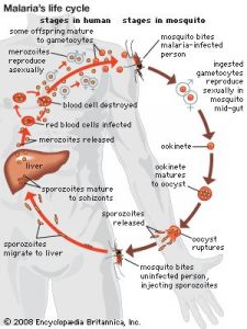
Mosquito transmission cycle

  • Uninfected mosquito. A mosquito becomes infected by feeding on a person who has malaria.
  • Transmission of the parasite. If this mosquito bites you in the future, it can transmit malaria parasites to you.
  • In the liver. Once the parasites enter your body, they travel to your liver, where some types can lie dormant for up to a year.
  • In the bloodstream. When the parasites mature, they leave the liver and infect red blood cells. This is when people usually develop malaria symptoms.
  • To the next person. If you are bitten by an uninfected mosquito at this point in the cycle, it will become infected with malaria parasites and can pass them on to other people it bites.

Other modes of transmission

Because the parasites that cause malaria affect red blood cells, people can also get malaria from exposure to infected blood, including:

  • From mother to unborn child
  • Through blood transfusions.
  • By sharing needles used to inject drugs

Risk factor’s

The biggest risk factor for developing malaria is living in or visiting areas where the disease is common. These include the tropical and subtropical regions of:

  • Sub-Saharan Africa
  • South and Southeast Asia
  • pacific islands
  • Central America and northern South America

The degree of risk depends on local malaria control, seasonal changes in malaria rates, and the precautions you take to avoid mosquito bites.

Risks of a more serious illness

People at higher risk for serious illness include:

  • toddlers and babies
  • Older adults
  • Travelers from malaria-free areas
  • Pregnant women and their unborn children

In many countries with high rates of malaria, the problem is compounded by a lack of access to preventive measures, medical care and information.

Immunity may decrease

Residents of a region with malaria may be exposed enough to the disease to acquire partial immunity, which can lessen the severity of malaria symptoms. However, this partial immunity can disappear if you move to a place where you are no longer frequently exposed to the parasite.

Complications

Malaria can be fatal, particularly when caused by Plasmodium species common in Africa. The World Health Organization estimates that around 94% of all malaria deaths occur in Africa, most commonly in children under 5 years of age. Malaria deaths are usually related to one or more serious complications, including:

  • Cerebral malaria. If parasite-filled blood cells block small blood vessels in the brain (cerebral malaria), brain swelling or brain damage can occur. Cerebral malaria can cause seizures and coma.
  • Respiratory problems. Fluid built up in the lungs (pulmonary oedema) can make it hard to breathe.
  • Organ failure. Malaria can damage the kidneys or liver or cause the spleen to rupture. Any of these conditions can be life-threatening.
  • Anaemia. Malaria can cause you do not to have enough red blood cells to adequately supply your body’s tissues with oxygen (anaemia).
  • Low blood sugar. Severe forms of malaria can cause low blood sugar (hypoglycemia), as can quinine, a common medicine used to combat malaria. Very low blood sugar can cause coma or death.

Malaria may reappear

Some varieties of the malaria parasite, which usually cause milder forms of the disease, can persist for years and cause relapses.

Prevention

If you live in or travel to an area where malaria is common, take steps to avoid mosquito bites. Mosquitoes are most active between dusk and dawn. To protect yourself from mosquito bites, you should:

  • Cover your skin. Wear pants and long-sleeved shirts. Put on your shirt and tuck your pant legs into your socks.
  • Apply insect repellent to the skin. Use an insect repellent registered with the Environmental Protection Agency on any exposed skin. These include repellents that contain DEET, picaridin, IR3535, oil of lemon eucalyptus (OLE), para-menthane-3,8-diol (PMD), or 2-undecanoate. Do not use a spray directly on your face. Do not use products with OLE or PMD in children under 3 years of age.
  • Apply repellent to clothing. Sprays containing permethrin are safe to apply to clothing.
  • Sleep under a net. Bed nets, particularly those treated with insecticides such as permethrin, help prevent mosquito bites while you sleep.

Preventive medicine

If you’re travelling to a place where malaria is common, talk to your doctor a few months beforehand about whether you should take medication before, during, and after your trip to help protect you from malaria parasites. In general, the medicines taken to prevent malaria are the same ones used to treat the disease. The medicine you take depends on where and how long you travel and on your own health.

Vaccine

The World Health Organization has recommended a malaria vaccine for use in children living in countries with a high number of malaria cases. Researchers continue to develop and study malaria vaccines to prevent infection.

Deficiency of Acyl-CoA Synthetase 5 (ACSL5) is Associated with a Severe and Treatable Failure to Thrive of Neonatal Onset

Deficiency of Acyl-CoA Synthetase 5 (ACSL5) is Associated with a Severe and Treatable Failure to Thrive of Neonatal Onset

Failure to thrive (FTT) causes important morbidity, usually with out clear etiologies. Six people of a giant consanguineous household introduced within the neonatal interval with recurrent vomiting and diarrhea, main to extreme FTT. Standard diagnostic work up didn’t verify an etiology.

Autozygosity mapping and complete exome sequencing recognized homozygosity for a novel genetic variant of the lengthy chain fatty acyl-CoA synthetase 5 (ACSL5) shared among the many affected people (NM_203379.1:c.1358C > A:p.[Thr453Lys]). Autosomal recessive genotype-phenotype segregation was confirmed by Sanger sequencing.

Functional in-vitro evaluation of the ACSL5 variant by immunofluorescence, western blotting and enzyme assay urged that Thr453Lys is a loss-of-function mutation with none remaining exercise. ACSL5 belongs to a vital enzyme household required for lipid metabolism and is identified to contribute the main exercise within the mouse gut.

Based on the operate of ACSL5 in intestinal lengthy chain fatty acid metabolism and the gastroenterological signs, affected people have been handled with complete parenteral vitamin or medium-chain triglyceride (MCT) based mostly components restricted in long-chain triglycerides. The sufferers responded properly and comply with up means that therapy is solely required throughout formative years.

The evaluation of the results of autozygosity, measured because the change of the imply worth of a trait amongst offspring of genetic family members, reveals the existence of directional dominance or overdominance. In this examine we detect proof of the impact of autozygosity in Four out of 13 cardiometabolic disease-associated traits utilizing information from greater than 10,000 sub-Saharan African people recruited from Ghana, Burkina Faso, Kenya and South Africa.

The impact of autozygosity on these phenotypes is discovered to be sex-related, with inbreeding having a important lowering impact in males however a important growing impact in ladies for a number of traits (physique mass index, subcutaneous adipose tissue, low-density lipoproteins and complete levels of cholesterol). Overall, the impact of inbreeding melancholy is extra intense in males.

Differential results of inbreeding melancholy are additionally noticed between examine websites with totally different night-light depth used as proxy for city improvement. These outcomes recommend a directional dominant genetic part mediated by environmental interactions and sex-specific variations in genetic structure for these traits within the Africa Wits-INDEPTH partnership for Genomic Studies (AWI-Gen) cohort.

Runs of homozygosity and evaluation of inbreeding melancholy

Pedigree info was historically used to assess inbreeding. Availability of high-density marker panels gives an alternate to assess inbreeding, notably within the presence of incomplete and error-prone pedigrees. Assessment of autozygosity throughout chromosomal segments utilizing runs of homozygosity (ROH) has emerged as a invaluable device to estimate inbreeding due to its common flexibility and means to quantify chromosomal contribution to genome-wide inbreeding.

Unfortunately, identification of ROH segments is delicate to the parameters used in the course of the search course of. These parameters are heuristically set, main to important variation within the outcomes. The minimal size required to determine a ROH section has main results on the estimation of inbreeding and inbreeding melancholy, but it is arbitrarily set.

To overcome this limitation, a search algorithm to approximate mutation enrichment was developed to decide the minimal size of ROH segments. It consists find genome segments with important impact variations in trait means between animals with excessive and low burdens of autozygous intervals with a particular size. The minimal size may very well be decided heuristically because the smallest interval at which a important sign is detected.

The proposed technique was examined in an inbred Hereford cattle inhabitants genotyped for 30,220 SNPs. Phenotypes recorded for six traits have been used for the approximation of mutation masses. The estimated minimal size was round 1 Mb for yearling weight (YW) and common every day acquire (ADG), and 4 Mb for delivery weight (BW) and weaning weight (WW).

These trait-specific thresholds estimated utilizing the proposed technique may very well be attributed to a trait-dependent impact of homozygosity. The detection of important inbreeding results was properly aligned with the estimated thresholds, particularly for YW and ADG. Although extremely deleterious alleles are anticipated to be extra frequent in latest inbreeding (lengthy ROH), brief ROH segments (< 5 Mb) may include a giant quantity of much less deleterious mutations with substantial joint results on some traits (YW and ADG).

Our outcomes spotlight the significance of correct estimation of the ROH-based inbreeding and the need to contemplate a trait-specific minimal size threshold for the identification of ROH segments in inbreeding melancholy analyses. These thresholds may very well be decided utilizing the proposed technique supplied the provision of phenotypic info.

 Deficiency of Acyl-CoA Synthetase 5 (ACSL5) is Associated with a Severe and Treatable Failure to Thrive of Neonatal Onset

Refining the genetic construction and relationships of European cattle breeds via meta-analysis of worldwide genomic SNP information, specializing in Italian cattle

The availability of genotyping assays has allowed the detailed analysis of cattle genetic range worldwide. However, these complete research didn’t embrace some native European populations, together with autochthonous Italian cattle. In this examine, we assembled a large-scale, genome-wide dataset of single nucleotide polymorphisms scored in 3,283 people from 205 cattle populations worldwide to assess genome-wide autozygosity and perceive higher the genetic relationships amongst these populations.

We prioritized European cattle, with a particular give attention to Italian breeds. Moderate variations in estimates of molecular inbreeding calculated from runs of homozygosity (FROH) have been noticed amongst domesticated bovid populations from totally different geographic areas, apart from Bali cattle.

Hepatitis B surface antigen Antibody

GWB-AA9974 0.5 mg Ask for price

Hepatitis B surface antigen Antibody

GWB-3250E0 0.1 mg Ask for price

Hepatitis B Surface Antigen Antibody

GWB-7EBDD9 0.1 ml Ask for price

Hepatitis B surface antigen Antibody

GWB-8B7121 0.5 mg Ask for price

Hepatitis B Surface Antigen Antibody

GWB-F21AB1 1 mg Ask for price

Hepatitis B surface antigen Antibody

GWB-F44C6A 0.1 mg Ask for price

Hepatitis B Surface Antigen Antibody

GWB-D8AC18 0.2 mg Ask for price

Hepatitis B Surface Antigen Antibody (HRP)

GWB-7F64AF 0.1 mg Ask for price

Hepatitis B Surface Antigen Antibody (Ad/Ay)

GWB-406C01 1 ml Ask for price

Hepatitis B Surface Antigen Antibody (Ad/Ay)

GWB-B67323 0.2 mg Ask for price

Hepatitis B Surface Antigen (HBsAg) Antibody

abx239778-100ug 100 ug
EUR 577.2

Hepatitis B Surface Antigen (HBsAg) Antibody

abx023848-1mg 1 mg
EUR 718.8

Hepatitis B Surface Antigen (HBsAg) Antibody

abx021777-1mg 1 mg
EUR 1220.4

Hepatitis B Surface Antigen (HBsAg) Antibody

abx021778-1mg 1 mg
EUR 1220.4

Hepatitis B Surface Antigen (HBsAg) Antibody

abx021779-1mg 1 mg
EUR 1220.4

Hepatitis B Surface Antigen (HBsAg) Antibody

abx021780-1mg 1 mg
EUR 1220.4

Hepatitis B Surface Antigen (HBsAg) Antibody

abx021781-1mg 1 mg
EUR 1178.4

Hepatitis B Surface Antigen (HBsAg) Antibody

abx021782-1mg 1 mg
EUR 718.8

Hepatitis B Surface Antigen (HBsAg) Antibody

abx021783-1mg 1 mg
EUR 718.8

Hepatitis B Surface Antigen (HBsAg) Antibody

abx021784-1mg 1 mg
EUR 718.8

Hepatitis B Surface Antigen (HBsAg) Antibody

abx021785-1mg 1 mg
EUR 718.8

Hepatitis B Surface Antigen (HBsAg) Antibody

abx021786-1mg 1 mg
EUR 718.8

Hepatitis B Surface Antigen (HBsAg) Antibody

abx021806-1mg 1 mg
EUR 718.8

Hepatitis B Surface Antigen (HBsAg) Antibody

abx021807-1mg 1 mg
EUR 718.8

Hepatitis B Surface Antigen (HBsAg) Antibody

abx021808-1mg 1 mg
EUR 718.8

Hepatitis B Surface Antigen (HBsAg) Antibody

abx021809-1mg 1 mg
EUR 718.8

Hepatitis B Surface Antigen (HBsAg) Antibody

abx021810-1mg 1 mg
EUR 718.8

Hepatitis B Surface Antigen (HBsAg) Antibody

abx021811-1mg 1 mg
EUR 777.6

Hepatitis B Surface Antigen (HBsAg) Antibody

abx021812-1mg 1 mg
EUR 718.8

Hepatitis B Surface Antigen (HBsAg) Antibody

abx021814-1mg 1 mg
EUR 1387.2

Hepatitis B Surface Antigen (HBsAg) Antibody

abx021815-1mg 1 mg
EUR 1387.2

Hepatitis B Surface Antigen (HBsAg) Antibody

abx021816-1mg 1 mg
EUR 718.8

Hepatitis B Surface Antigen (HBsAg) Antibody

abx021817-100ug 100 ug
EUR 794.4

Hepatitis B Surface Antigen (HBsAg) Antibody

abx021818-1mg 1 mg
EUR 777.6

Hepatitis B Surface Antigen (HBsAg) Antibody

abx021819-1mg 1 mg
EUR 661.2

Hepatitis B Surface Antigen (HBsAg) Antibody

abx021820-1mg 1 mg
EUR 661.2

Hepatitis B Surface Antigen (HBsAg) Antibody

abx021821-1mg 1 mg
EUR 661.2

Hepatitis B Surface Antigen (HBsAg) Antibody

abx021823-1mg 1 mg
EUR 661.2

Hepatitis B Surface Antigen (HBsAg) Antibody

abx022999-1mg 1 mg
EUR 577.2

Hepatitis B Surface Antigen (HBsAg) Antibody

abx023004-1mg 1 mg
EUR 811.2

Hepatitis B Surface Antigen (HBsAg) Antibody

abx023006-1ml 1 ml
EUR 777.6

Hepatitis B Surface Antigen (HBsAg) Antibody

abx023007-1ml 1 ml
EUR 718.8

Hepatitis B Surface Antigen (HBsAg) Antibody

abx021777-400l 400 µl Ask for price

Hepatitis B Surface Antigen (HBsAg) Antibody

abx021777-80l 80 µl
EUR 1175

Hepatitis B Surface Antigen (HBsAg) Antibody

abx021778-400l 400 µl Ask for price

Hepatitis B Surface Antigen (HBsAg) Antibody

abx021778-80l 80 µl
EUR 1175

Hepatitis B Surface Antigen (HBsAg) Antibody

abx021779-400l 400 µl Ask for price

Hepatitis B Surface Antigen (HBsAg) Antibody

abx021779-80l 80 µl
EUR 1175

Hepatitis B Surface Antigen (HBsAg) Antibody

abx021780-400l 400 µl Ask for price

Hepatitis B Surface Antigen (HBsAg) Antibody

abx021780-80l 80 µl
EUR 1175

Hepatitis B Surface Antigen (HBsAg) Antibody

abx021781-400l 400 µl Ask for price

Hepatitis B Surface Antigen (HBsAg) Antibody

abx021781-80l 80 µl
EUR 1175

Hepatitis B Surface Antigen (HBsAg) Antibody

abx021782-400l 400 µl Ask for price

Hepatitis B Surface Antigen (HBsAg) Antibody

abx021782-80l 80 µl
EUR 787.5

Hepatitis B Surface Antigen (HBsAg) Antibody

abx021783-400l 400 µl Ask for price

Hepatitis B Surface Antigen (HBsAg) Antibody

abx021783-80l 80 µl
EUR 787.5

Hepatitis B Surface Antigen (HBsAg) Antibody

abx021784-400l 400 µl Ask for price

Hepatitis B Surface Antigen (HBsAg) Antibody

abx021784-80l 80 µl
EUR 787.5

Hepatitis B Surface Antigen (HBsAg) Antibody

abx021785-400l 400 µl Ask for price

Hepatitis B Surface Antigen (HBsAg) Antibody

abx021785-80l 80 µl
EUR 787.5

Hepatitis B Surface Antigen (HBsAg) Antibody

abx021786-400l 400 µl Ask for price

Hepatitis B Surface Antigen (HBsAg) Antibody

abx021786-80l 80 µl
EUR 725

Hepatitis B Surface Antigen (HBsAg) Antibody

abx021806-400l 400 µl Ask for price

Hepatitis B Surface Antigen (HBsAg) Antibody

abx021806-80l 80 µl
EUR 687.5

Hepatitis B Surface Antigen (HBsAg) Antibody

abx021807-400l 400 µl Ask for price

Hepatitis B Surface Antigen (HBsAg) Antibody

abx021807-80l 80 µl
EUR 687.5

Hepatitis B Surface Antigen (HBsAg) Antibody

abx021809-400l 400 µl Ask for price

Hepatitis B Surface Antigen (HBsAg) Antibody

abx021809-80l 80 µl
EUR 662.5

Hepatitis B Surface Antigen (HBsAg) Antibody

abx021810-400l 400 µl Ask for price

Hepatitis B Surface Antigen (HBsAg) Antibody

abx021810-80l 80 µl
EUR 762.5

Hepatitis B Surface Antigen (HBsAg) Antibody

abx021811-400l 400 µl Ask for price

Hepatitis B Surface Antigen (HBsAg) Antibody

abx021811-80l 80 µl
EUR 825

Hepatitis B Surface Antigen (HBsAg) Antibody

abx021812-400l 400 µl Ask for price

Hepatitis B Surface Antigen (HBsAg) Antibody

abx021812-80l 80 µl
EUR 762.5

Hepatitis B Surface Antigen (HBsAg) Antibody

abx021814-400l 400 µl Ask for price

Hepatitis B Surface Antigen (HBsAg) Antibody

abx021814-80l 80 µl
EUR 375

Hepatitis B Surface Antigen (HBsAg) Antibody

abx021815-400l 400 µl Ask for price

Hepatitis B Surface Antigen (HBsAg) Antibody

abx021815-80l 80 µl
EUR 1175

Hepatitis B Surface Antigen (HBsAg) Antibody

abx021816-400l 400 µl Ask for price

Hepatitis B Surface Antigen (HBsAg) Antibody

abx021816-80l 80 µl
EUR 687.5

Hepatitis B Surface Antigen (HBsAg) Antibody

abx021817-400l 400 µl Ask for price

Hepatitis B Surface Antigen (HBsAg) Antibody

abx021817-80l 80 µl
EUR 725

Hepatitis B Surface Antigen (HBsAg) Antibody

abx021818-400l 400 µl Ask for price

Hepatitis B Surface Antigen (HBsAg) Antibody

abx021818-80l 80 µl
EUR 825

Hepatitis B Surface Antigen (HBsAg) Antibody

abx021821-400l 400 µl Ask for price

Hepatitis B Surface Antigen (HBsAg) Antibody

abx021821-80l 80 µl
EUR 637.5

Hepatitis B Surface Antigen (HBsAg) Antibody

abx023006-400l 400 µl Ask for price

Hepatitis B Surface Antigen (HBsAg) Antibody

abx023006-80l 80 µl
EUR 1050

Hepatitis B Surface Antigen (HBsAg) Antibody

abx023007-400l 400 µl Ask for price

Hepatitis B Surface Antigen (HBsAg) Antibody

abx023007-80l 80 µl
EUR 975

Hepatitis B Surface Antigen (HBsAg) Antibody

abx239778-100l 100 µl Ask for price

Hepatitis B Surface Antigen (HBsAg) Antibody

abx239778-50l 50 µl
EUR 387.5

Hepatitis B Surface Antigen (HBsAg) Antibody

abx240304-100l 100 µl Ask for price

Hepatitis B Surface Antigen (HBsAg) Antibody

abx240304-50l 50 µl
EUR 350

Hepatitis B Surface Antigen (HBsAg) Antibody

abx376359-96tests 96 tests
EUR 225

Hepatitis B Surface Antigen (HBsAg) Antibody

abx342052-100l 100 µl Ask for price

Hepatitis B Surface Antigen (HBsAg) Antibody

abx342052-50l 50 µl
EUR 2575

Hepatitis B Surface Antigen (HBsAg) Antibody

abx342091-100l 100 µl Ask for price

Hepatitis B Surface Antigen (HBsAg) Antibody

abx342091-50l 50 µl
EUR 2575

Hepatitis B Virus surface antigen Antibody

GWB-4BAE14 1 ml Ask for price

Hepatitis B Virus Surface Antigen Antibody

20-abx134235
  • Ask for price
  • Ask for price
  • Ask for price
  • 100 ul
  • 200 ul
  • 30 ul

Hepatitis B Virus Surface Antigen Antibody

20-abx134236
  • Ask for price
  • Ask for price
  • Ask for price
  • 100 ul
  • 200 ul
  • 30 ul

HEPATITIS B SURFACE ANTIGEN AY

GWB-F1CE5F 0.1 mg Ask for price

Hepatitis A Surface Antigen Antibody

GWB-DFB8F4 0.2 mg Ask for price

Hepatitis A Surface Antigen Antibody

GWB-8A0702 0.2 mg Ask for price

Hepatitis B Surface Antigen Antibody (AD/AY) (FITC)

GWB-ABE5A1 1 ml Ask for price

Hepatitis B Surface Antigen (HBsAg) Antibody (HRP)

abx021776-1mg 1 mg
EUR 1296

Hepatitis B Surface Antigen (HBsAg) Antibody (HRP)

abx021822-1mg 1 mg
EUR 1296

Hepatitis B Surface Antigen (HBsAg) Antibody (HRP)

abx023002-1ml 1 ml
EUR 718.8

Hepatitis B Surface Antigen (HBsAg) Antibody (HRP)

abx023005-1ml 1 ml
EUR 886.8

Hepatitis B Surface Antigen (HBsAg) Antibody (HRP)

abx021776-400l 400 µl Ask for price

Hepatitis B Surface Antigen (HBsAg) Antibody (HRP)

abx021776-80l 80 µl
EUR 1175

Hepatitis B Surface Antigen (HBsAg) Antibody (HRP)

abx023002-400l 400 µl Ask for price

Hepatitis B Surface Antigen (HBsAg) Antibody (HRP)

abx023002-80l 80 µl
EUR 1112.5

Hepatitis B Surface Antigen (HBsAg) Antibody (HRP)

abx023005-400l 400 µl Ask for price

Hepatitis B Surface Antigen (HBsAg) Antibody (HRP)

abx023005-80l 80 µl
EUR 1175

Hepatitis B Surface Antigen (HBsAg) Antibody (FITC)

abx023001-1ml 1 ml
EUR 718.8

Hepatitis B Surface Antigen (HBsAg) Antibody (FITC)

abx023009-1ml 1 ml
EUR 844.8

Hepatitis B Surface Antigen (HBsAg) Antibody Pair

abx137761-100l 100 µl
EUR 350

Hepatitis B Surface Antigen (HBsAg) Antibody Pair

abx137761-500l 500 µl
EUR 950

Hepatitis B Surface Antigen (HBsAg) Antibody (FITC)

abx023001-400l 400 µl Ask for price

Hepatitis B Surface Antigen (HBsAg) Antibody (FITC)

abx023001-80l 80 µl
EUR 1112.5

Hepatitis B Surface Antigen (HBsAg) Antibody (FITC)

abx023009-400l 400 µl Ask for price

Hepatitis B Surface Antigen (HBsAg) Antibody (FITC)

abx023009-80l 80 µl
EUR 1175

Hepatitis B Surface Antigen Antibody (ready to use)

GWB-95C198 6 ml Ask for price

Hepatitis B Virus Surface Antigen Antibody (HRP)

GWB-475DB7 0.1 ml Ask for price

Hepatitis B Virus Surface Antigen Antibody (HRP)

20-abx134237
  • Ask for price
  • Ask for price
  • Ask for price
  • 100 ul
  • 200 ul
  • 30 ul

Hepatitis B Virus Surface Antigen Antibody (HRP)

20-abx134238
  • Ask for price
  • Ask for price
  • Ask for price
  • 100 ul
  • 200 ul
  • 30 ul

OASA00809-200UG - HEPATITIS B SURFACE ANTIGEN Antibody

OASA00809-200UG 0.2mg
EUR 459

OASA00814-100UG - HEPATITIS B SURFACE ANTIGEN Antibody

OASA00814-100UG 0.1mg
EUR 469

Hepatitis B Surface Antigen (HBsAg) Antibody (Biotin)

abx023000-1ml 1 ml
EUR 718.8

Hepatitis B Surface Antigen (HBsAg) Antibody (Biotin)

abx023008-1ml 1 ml
EUR 844.8

Hepatitis B Surface Antigen (HBsAg) Antibody (Biotin)

abx023000-400l 400 µl Ask for price

Hepatitis B Surface Antigen (HBsAg) Antibody (Biotin)

abx023000-80l 80 µl
EUR 1050

Hepatitis B Surface Antigen (HBsAg) Antibody (Biotin)

abx023008-400l 400 µl Ask for price

Hepatitis B Surface Antigen (HBsAg) Antibody (Biotin)

abx023008-80l 80 µl
EUR 1087.5

Hepatitis B Virus Surface Antigen Antibody (FITC)

20-abx134239
  • Ask for price
  • Ask for price
  • Ask for price
  • 100 ul
  • 200 ul
  • 30 ul

Hepatitis B Virus Surface Antigen Antibody (FITC)

20-abx134240
  • Ask for price
  • Ask for price
  • Ask for price
  • 100 ul
  • 200 ul
  • 30 ul

Hepatitis B Virus Surface Antigen Antibody (Biotin)

GWB-13CFC6 0.1 ml Ask for price

Hepatitis B Virus Surface Antigen Antibody (Biotin)

20-abx134241
  • Ask for price
  • Ask for price
  • Ask for price
  • 100 ul
  • 200 ul
  • 30 ul

Hepatitis B Virus Surface Antigen Antibody (Biotin)

20-abx134242
  • Ask for price
  • Ask for price
  • Ask for price
  • 100 ul
  • 200 ul
  • 30 ul

Hepatitis B Virus Surface Antigen Antibody (Biotin)

GWB-A855CE 0.1 ml Ask for price

Hepatitis B Surface Antigen (HBsAg) Antibody (ad/ay)

GWB-T00719 1 mg Ask for price

Recombinant Hepatitis B Surface Antigen

DAG435 100ug
EUR 878.4

Hepatitis B Antibody (Virus Surface Antigen) (FITC)

GWB-8ABE07 0.1 ml Ask for price

Hepatitis B,surface Antigen, (HbsAg) Antibody, Concentrate

PAB361C 0.2ml
EUR 455

Hepatitis B Virus Surface Antigen (HBsAg) Antibody

20-abx137498
  • Ask for price
  • Ask for price
  • Ask for price
  • 10 ug
  • 1 mg
  • 2 µg

Hepatitis B Virus Surface Antigen (HBsAg) Antibody

abx137498-100tests 100 tests
EUR 225

Hepatitis B Surface Antigen Rabbit mAb

E2R50191 100ul
EUR 385
Description: Biotin-Conjugated, FITC-Conjugated , AF350 Conjugated , AF405M-Conjugated ,AF488-Conjugated, AF514-Conjugated ,AF532-Conjugated, AF555-Conjugated ,AF568-Conjugated , HRP-Conjugated, AF405S-Conjugated, AF405L-Conjugated , AF546-Conjugated, AF594-Conjugated , AF610-Conjugated, AF635-Conjugated , AF647-Conjugated , AF680-Conjugated , AF700-Conjugated , AF750-Conjugated , AF790-Conjugated , APC-Conjugated , PE-Conjugated , Cy3-Conjugated , Cy5-Conjugated , Cy5.5-Conjugated , Cy7-Conjugated Antibody

ELISA Kit for Antibody to Hepatitis B Surface Antigen

KO31004096 1 kit
EUR 247

Hepatitis B Surface Antigen preS1 Protein

20-abx260483
  • Ask for price
  • Ask for price
  • Ask for price
  • 10 ug
  • 1 mg
  • 50 ug

Hepatitis B Surface Antigen preS2 Protein

20-abx260485
  • Ask for price
  • Ask for price
  • Ask for price
  • 10 ug
  • 1 mg
  • 50 ug

Hepatitis B Surface Antigen preS2 Protein

abx260485-10g 10 µg
EUR 325

Hepatitis B Surface Antigen preS2 Protein

abx260485-2g 2 µg
EUR 212.5

Hepatitis B Surface Antigen (HBsAg) Protein

abx060552-100g 100 µg Ask for price

Hepatitis B Surface Antigen (HBsAg) Protein

abx060552-10g 10 µg
EUR 1362.5

Hepatitis B Surface Antigen (HBsAg) Protein

abx060552-50g 50 µg Ask for price

RECOMBINANT HEPATITIS B SURFACE ANTIGEN AD

GWB-BEC112 0.1 mg Ask for price

Mouse Anti Hepatitis B Surface Antigen Monoclonal Antibody

DMABT-51323MH 0.2 mg
EUR 696

Mouse Anti Hepatitis B Surface Antigen Monoclonal Antibody

DMABT-51324MH 0.1 ml
EUR 534

Mouse Anti Hepatitis B Surface Antigen Monoclonal Antibody

DMABT-51325MH 1 ml
EUR 982.8

Mouse Anti Hepatitis B Surface Antigen Monoclonal Antibody

DMABT-51326MH 1 ml
EUR 534

Mouse Anti Hepatitis B Surface Antigen Monoclonal Antibody

DMABT-51327MH 6 ml
EUR 858

Mouse Anti Hepatitis B Surface Antigen Monoclonal Antibody

DMABT-51328MH 0.1 mg
EUR 889.2

Hepatitis B Surface Antigen, ayw Recombinant

rAP-5201 Inquiry Ask for price

Hepatitis B Surface Antigen, adw Recombinant

rAP-5203 Inquiry Ask for price

Hepatitis B Surface Antigen ayw Recombinant

rAP-5207 Inquiry Ask for price

OAEF00317-1MG - Hepatitis B Surface Antigen (HBsAg) Antibody

OAEF00317-1MG 1mg
EUR 236

OAEF00318-1MG - Hepatitis B Surface Antigen (HBsAg) Antibody

OAEF00318-1MG 1mg
EUR 236

OAEF00351-1MG - Hepatitis B Surface Antigen (HBsAg) Antibody

OAEF00351-1MG 1mg
EUR 197

OAEF00404-1MG - Hepatitis B Surface Antigen (HBsAg) Antibody

OAEF00404-1MG 1mg
EUR 229

OAEF00405-1MG - Hepatitis B Surface Antigen (HBsAg) Antibody

OAEF00405-1MG 1mg
EUR 229

OAEF00918-1MG - Hepatitis B Surface Antigen (HBsAg) Antibody

OAEF00918-1MG 1mg
EUR 236

OAEF00919-1MG - Hepatitis B Surface Antigen (HBsAg) Antibody

OAEF00919-1MG 1mg
EUR 236

OAEF00920-1MG - Hepatitis B Surface Antigen (HBsAg) Antibody

OAEF00920-1MG 1mg
EUR 236

OAEF00975-1MG - Hepatitis B Surface Antigen (HBsAg) Antibody

OAEF00975-1MG 1mg
EUR 439

Our findings indicated that some Italian breeds present the very best estimates of ranges of molecular inbreeding among the many cattle populations assessed on this examine. Patterns of genetic differentiation, shared ancestry, and phylogenetic evaluation all urged the incidence of gene circulate, notably amongst populations originating from the identical geographical space.

For European cattle, we noticed a distribution alongside three fundamental instructions, reflecting the identified historical past and formation of the analyzed breeds. The Italian breeds are cut up into two fundamental teams, based mostly on their historic origin and diploma of conservation of ancestral genomic parts. The outcomes pinpointed that additionally Sicilian breeds, a lot alike Podolian derived-breeds, up to now skilled a related non-European affect, with African and indicine introgression.

Genome-Wide Assessment of Runs of Homozygosity in Chinese Wagyu Beef Cattle

Genome-Wide Assessment of Runs of Homozygosity in Chinese Wagyu Beef Cattle

Runs of homozygosity (ROH) are steady homozygous areas that usually exist in the DNA sequence of diploid organisms. Identifications of ROH resulting in discount in efficiency can present priceless perception into the genetic structure of advanced traits. Here, we evaluated genome-wide patterns of homozygosity and their affiliation with essential traits in Chinese Wagyu beef cattle.

We recognized a complete of 29,271 ROH segments from 462 animals. Within every animal, a median quantity of ROH was 63.36 whereas a median size was 62.19 Mb. To consider the enrichment of ROH throughout genomes, we initially recognized 280 ROH areas by merging ROH occasions throughout all people.

Of these, 9 areas containing 154 candidate genes, had been considerably related to six traits (physique peak, chest circumference, fats protection, backfat thickness, ribeye space, and carcass size; p < 0.01). Moreover, we discovered 26 consensus ROH areas with frequencies exceeding 10%, and a number of other areas overlapped with QTLs, that are related to physique weight, calving ease, and stillbirth. Among them, we noticed 41 candidate genes, together BCKDHBMAB21L1SLC2A13FGFR3FGFRL1CPLX1CTNNA1CORTCTNNBIP1, and NMNAT1, which have been beforehand reported to be associated to physique conformation, meat high quality, susceptibility, and reproductive traits. In abstract, we assessed genome-wide autozygosity patterns and inbreeding ranges in Chinese Wagyu beef cattle. Our examine recognized many candidate areas and genes overlapped with ROH for a number of essential traits, which could possibly be unitized to help the design of a variety mating technique in beef cattle.

The genomic variability of native Italian rooster breeds, which had been monitored beneath a conservation plan, was studied utilizing single nucleotide polymorphisms (SNPs) to grasp their genetic range and inhabitants construction. A complete of 582 samples from 23 native breeds and 4 business shares had been genotyped utilizing the Affymetrix 600 Ok Chicken SNP Array. In common, the degrees of genetic range, investigated by way of completely different approaches, had been lowest in the native rooster breeds in comparison with these in the business shares.

The stage of genomic inbreeding, primarily based on runs of homozygosity (FROH), was markedly completely different among the many breeds and ranged from 0.121 (Valdarnese) to 0.607 (Siciliana). In all breeds, brief runs of homozygosity (ROH) (<4 Mb in size) had been extra frequent than lengthy segments.

The patterns of genetic differentiation, model-based clustering, and neighbor networks confirmed that the majority breeds shaped non-overlapping clusters and had been clearly separate populations, which indicated the presence of gene move, particularly amongst breeds that originated from the identical geographical space.

Genetic spectrum of retinal dystrophies in Tunisia

We report the molecular foundation of the biggest Tunisian cohort with inherited retinal dystrophies (IRD) reported to this point, establish disease-causing pathogenic variants and describe genotype-phenotype correlations. A subset of 26 households from a cohort of 73 households with scientific analysis of autosomal recessive IRD (AR-IRD) excluding Usher syndrome was analyzed by entire exome sequencing and autozygosity mapping.

Causative pathogenic variants had been recognized in 50 households (68.4%), 42% of which had been novel. The most prevalent pathogenic variants had been noticed in ABCA4 (14%) and RPE65, CRB1 and CERKL (8% every). 26 variants (Eight novel and 18 recognized) in 19 genes had been recognized in 26 households (14 missense substitutions, 5 deletions, Four nonsense pathogenic variants and three splice website variants), with additional allelic heterogeneity arising from completely different pathogenic variants in the identical gene.

The most typical phenotype in our cohort is retinitis pigmentosa (23%) and cone rod dystrophy (23%) adopted by Leber congenital amaurosis (19.2%). We report the affiliation of new illness phenotypes. This analysis was carried out in Tunisian sufferers with IRD in order to delineate the genetic inhabitants structure.

Genome-Wide Assessment of Runs of Homozygosity in Chinese Wagyu Beef Cattle

Shadow autozygosity mapping by linkage exclusion (SAMPLE): a easy technique to establish the genetic foundation of deadly autosomal recessive problems.

Autozygosity mapping has been invaluable for figuring out the genetic foundation of deadly autosomal recessive problems, however this strategy stays difficult as a result of DNA from affected people might typically be unavailable or of inadequate high quality for intensive molecular genetic research. To circumvent these difficulties, we developed a pc program known as “SAMPLE” (for shadow autozygosity mapping by linkage exclusion) to reinforce autozygosity mapping by way of the empirical evaluation of haplotypes of unaffected people in consanguineous households.

Single nucleotide polymorphism (SNP) genotyping of unaffected people in advanced consanguineous pedigrees is used to deduce restricted chromosomal areas suitable with linkage to a possible illness locus, and to permit the instant prioritization of potential areas of curiosity. Further restricted genotyping then permits the speedy affirmation and advantageous mapping of a illness locus.

We display the utility of this technique by utilizing genotyping information from solely mother and father and unaffected siblings, in three consanguineous households affected with Meckel-Gruber syndrome, to accurately infer the situation of the MKS3/TMEM67 locus on chromosome 8q22.1.

Recombinant Mouse Prostate stem cell antigen (Psca)

CSB-YP018840MO 5939 mg Ask for price

Recombinant Mouse Prostate stem cell antigen(Psca)

AP72740 each Ask for price

Recombinant Mouse Prostate stem cell antigen (Psca)

RPC20241-100ug 100ug
EUR 569.8

Recombinant Mouse Prostate stem cell antigen (Psca)

RPC20241-20ug 20ug
EUR 353.4

StemTAG PCR Primer Set for Stem Cell Characterization

CBA-303 1 kit
EUR 470.4
Description: StemTAG PCR Primer Set for Stem Cell Characterization includes 7 primer pairs: Oct-4, NANOG, AFP, Flk-1, and NCAM, plus GAPDH and beta-actin as controls.

Prostate stem cell antigen

AP86675 1mg
EUR 2640

Prostate stem cell antigen

AP87071 1mg
EUR 2640

Mouse Psca/ Prostate stem cell antigen ELISA Kit

E1215Mo 1 Kit
EUR 543

Mouse Prostate stem cell antigen, Psca ELISA KIT

ELI-22375m 96 Tests
EUR 1038

Mouse Prostate stem cell antigen (PSCA) ELISA Kit

abx520719-96tests 96 tests
EUR 886.8

Mouse Prostate stem cell antigen (PSCA) ELISA Kit

RK13577 96T
EUR 280

Mouse Psca / Prostate stem cell antigen ELISA Kit

E8693m 96T
EUR 776

Total Protein - Murine Embryonic Stem Cell Line D3

CBA-305 500 ?g
EUR 414
Description:
  • Isolated from mouse ES-D3 cell line
  • Presented as 500 µg at 1 mg/mL in NP-40 Solubilization Buffer

CytoSelect Proliferating Cell Nuclear Antigen (PCNA) ELISA Kit

CBA-254 96 assays
EUR 672
Description: Cell Biolabs? CytoSelect Proliferating Cell Nuclear Antigen (PCNA) ELISA Kit is an enzyme immunoassay developed for the detection and quantitation of PCNA from nuclear and whole cell extracts.  The kit detects PCNA from mouse, rat and human, and has a detection sensitivity limit of 12.5 ng/mLPCNA.  Each kit provides sufficient reagents to perform up to 96 assays including standard curve and unknown samples. 

Prostate Stem Cell Antigen Antibody

33476 100ul
EUR 319

Prostate Stem Cell Antigen Antibody

33476-100ul 100ul
EUR 302.4

Prostate Stem Cell Antigen Antibody

33476-50ul 50ul
EUR 224.4

Prostate Stem Cell Antigen Antibody

E033476 100μg/100μl
EUR 255
Description: Available in various conjugation types.

Prostate Stem Cell Antigen Antibody

E19-8709-1 50ug/50ul
EUR 145
Description: Available in various conjugation types.

Prostate Stem Cell Antigen Antibody

E19-8709-2 100ug/100ul
EUR 225
Description: Available in various conjugation types.

Prostate Stem Cell Antigen Antibody

E11-0310C 100μg/100μl
EUR 225
Description: Available in various conjugation types.

Prostate Stem Cell Antigen Antibody

DF8709 200ul
EUR 420

Prostate Stem Cell Antigen Antibody

DF8709-100ul 100ul
EUR 280

Prostate Stem Cell Antigen Antibody

DF8709-200ul 200ul
EUR 350

Prostate Stem Cell Antigen Antibody

ABD8709 100 ug
EUR 525.6

Prostate Stem Cell Antigen Antibody

8C0310 50ug
EUR 368
Description: Prostate Stem Cell Antigen Antibody

OPCA01291-20UG - Recombinant Mouse Prostate stem cell antigen

OPCA01291-20UG 20ug
EUR 315

Psca (untagged) - Mouse prostate stem cell antigen (Psca), (10ug)

MC211324 10 µg Ask for price

OPCA01291-100UG - Recombinant Mouse Prostate stem cell antigen

OPCA01291-100UG 100ug
EUR 462

OPCA01291-500UG - Recombinant Mouse Prostate stem cell antigen

OPCA01291-500UG 500ug
EUR 1276

Psca (GFP-tagged) - Mouse prostate stem cell antigen (Psca), (10ug)

MG215317 10 µg Ask for price

Psca (Myc-DDK-tagged) - Mouse prostate stem cell antigen (Psca)

MR215317 10 µg Ask for price

Prostate stem cell antigen (PSCA) Antibody

20-abx325516
  • Ask for price
  • Ask for price
  • 100 ug
  • 50 ug

Prostate Stem Cell Antigen (PSCA) Antibody

abx433189-200ul 200 ul
EUR 343.2

Prostate Stem Cell Antigen (PSCA) Antibody

20-abx320478
  • Ask for price
  • Ask for price
  • 100 ul
  • 50 ul

Prostate stem cell antigen (PSCA) Antibody

abx331721-100ul 100 ul
EUR 510

Prostate stem cell antigen (PSCA) Antibody

20-abx211961
  • Ask for price
  • Ask for price
  • 100 ul
  • 50 ul

Prostate stem cell antigen (PSCA) Antibody

20-abx212351
  • Ask for price
  • Ask for price
  • 100 ul
  • 50 ul

Prostate Stem Cell Antigen (PSCA) Antibody

abx236848-100ug 100 ug
EUR 661.2

Prostate Stem Cell Antigen (PSCA) Antibody

20-abx114737
  • Ask for price
  • Ask for price
  • 150 ul
  • 50 ul

Prostate Stem Cell Antigen (PSCA) Antibody

20-abx102687
  • Ask for price
  • Ask for price
  • Ask for price
  • Ask for price
  • Ask for price
  • 100 ug
  • 10 ug
  • 1 mg
  • 200 ug
  • 50 ug

Prostate Stem Cell Antigen (PSCA) Antibody

20-abx102688
  • Ask for price
  • Ask for price
  • Ask for price
  • Ask for price
  • Ask for price
  • 100 ug
  • 10 ug
  • 1 mg
  • 200 ug
  • 50 ug

Prostate Stem Cell Antigen (PSCA) Antibody

20-abx132387
  • Ask for price
  • Ask for price
  • Ask for price
  • Ask for price
  • Ask for price
  • 100 ug
  • 1 mg
  • 200 ug
  • 20 ug
  • 50 ug

Prostate Stem Cell Antigen (PSCA) Antibody

20-abx008545
  • Ask for price
  • Ask for price
  • Ask for price
  • 100 ul
  • 200 ul
  • 30 ul

Prostate Stem Cell Antigen (PSCA) Antibody

20-abx013183
  • Ask for price
  • Ask for price
  • Ask for price
  • Ask for price
  • 100 ug
  • 10 ug
  • 200 ug
  • 300 µg

Prostate Stem Cell Antigen (PSCA) Antibody

abx027872-400ul 400 ul
EUR 627.6

Prostate Stem Cell Antigen (PSCA) Antibody

abx027872-80l 80 µl
EUR 343.2

Prostate Stem Cell Antigen (PSCA), Antibody

GWB-B3D35D 7 ml Ask for price

Prostate Stem Cell Antigen (PSCA), Antibody

GWB-983A18 1 ml Ask for price

Prostate Stem Cell Antigen (PSCA) Antibody

abx004291-100l 100 µl
EUR 400

Prostate Stem Cell Antigen (PSCA) Antibody

abx004291-20l 20 µl
EUR 175

Prostate Stem Cell Antigen (PSCA) Antibody

abx004291-50l 50 µl
EUR 275

Prostate Stem Cell Antigen (PSCA) Antibody

abx013183-100g 100 µg
EUR 237.5

Prostate Stem Cell Antigen (PSCA) Antibody

abx013183-10g 10 µg
EUR 43.75

Prostate Stem Cell Antigen (PSCA) Antibody

abx013183-200g 200 µg
EUR 312.5

Prostate Stem Cell Antigen (PSCA) Antibody

abx236848-100g 100 µg
EUR 350

Prostate Stem Cell Antigen (PSCA) Antibody

abx114737-100l 100 µl
EUR 612.5

Prostate Stem Cell Antigen (PSCA) Antibody

abx132387-01mg 0.1 mg
EUR 275

Prostate Stem Cell Antigen (PSCA) Antibody

abx102687-100l 100 µl
EUR 275

Prostate Stem Cell Antigen (PSCA) Antibody

abx102687-1ml 1 ml
EUR 750

Prostate Stem Cell Antigen (PSCA) Antibody

abx102687-200l 200 µl
EUR 337.5

Prostate Stem Cell Antigen (PSCA) Antibody

abx102688-100l 100 µl
EUR 275

Prostate Stem Cell Antigen (PSCA) Antibody

abx102688-1ml 1 ml
EUR 750

Prostate Stem Cell Antigen (PSCA) Antibody

abx102688-200l 200 µl
EUR 337.5

Prostate Stem Cell Antigen (PSCA) Antibody

abx027872-400l 400 µl
EUR 518.75

Prostate stem cell antigen (PSCA) Antibody

abx211961-100l 100 µl
EUR 350

Prostate stem cell antigen (PSCA) Antibody

abx211961-50l 50 µl
EUR 250

Prostate stem cell antigen (PSCA) Antibody

abx212351-100l 100 µl
EUR 350

Prostate stem cell antigen (PSCA) Antibody

abx212351-50l 50 µl
EUR 250

Prostate Stem Cell Antigen (PSCA) Antibody

abx320478-100l 100 µl
EUR 350

Prostate Stem Cell Antigen (PSCA) Antibody

abx320478-50l 50 µl
EUR 237.5

Prostate stem cell antigen (PSCA) Antibody

abx325516-100g 100 µg
EUR 250

Prostate stem cell antigen (PSCA) Antibody

abx325516-50g 50 µg
EUR 187.5

Prostate Stem Cell Antigen (PSCA) Antibody

abx433189-100g 100 µg
EUR 387.5

Human Prostate stem cell antigen (PSCA)

1-CSB-YP018840HU
  • Ask for price
  • Ask for price
  • Ask for price
  • Ask for price
  • Ask for price
  • Ask for price
  • 100ug
  • 10ug
  • 1MG
  • 200ug
  • 500ug
  • 50ug
Description: Recombinant Human Prostate stem cell antigen(PSCA) expressed in Yeast

Human Prostate stem cell antigen (PSCA)

1-CSB-EP018840HU
  • Ask for price
  • Ask for price
  • Ask for price
  • Ask for price
  • Ask for price
  • Ask for price
  • 100ug
  • 10ug
  • 1MG
  • 200ug
  • 500ug
  • 50ug
Description: Recombinant Human Prostate stem cell antigen(PSCA) expressed in E.coli

Human Prostate stem cell antigen (PSCA)

1-CSB-EP018840HUa0
  • Ask for price
  • Ask for price
  • Ask for price
  • Ask for price
  • Ask for price
  • Ask for price
  • 100ug
  • 10ug
  • 1MG
  • 200ug
  • 500ug
  • 50ug
Description: Recombinant Human Prostate stem cell antigen(PSCA) expressed in E.coli

Recombinant Mouse Prostate stem cell antigen Protein, His, Yeast-1mg

QP9353-ye-1mg 1mg
EUR 2763.6

Prostate Stem Cell Antigen Conjugated Antibody

C33476 100ul
EUR 476.4

Recombinant Mouse Prostate stem cell antigen Protein, His, Yeast-100ug

QP9353-ye-100ug 100ug
EUR 685.2

Recombinant Mouse Prostate stem cell antigen Protein, His, Yeast-10ug

QP9353-ye-10ug 10ug
EUR 326.4

Recombinant Mouse Prostate stem cell antigen Protein, His, Yeast-200ug

QP9353-ye-200ug 200ug
EUR 1077.6

Recombinant Mouse Prostate stem cell antigen Protein, His, Yeast-500ug

QP9353-ye-500ug 500ug
EUR 1806

Recombinant Mouse Prostate stem cell antigen Protein, His, Yeast-50ug

QP9353-ye-50ug 50ug
EUR 424.8

Prostate Stem Cell Antigen Blocking Peptide

DF8709-BP 1mg
EUR 234

Recombinant Prostate Stem Cell Antigen (PSCA)

4-RPC735Hu01
  • Ask for price
  • Ask for price
  • Ask for price
  • Ask for price
  • Ask for price
  • Ask for price
  • Ask for price
  • 100 ug
  • 10ug
  • 1 mg
  • 200 ug
  • 500 ug
  • 50ug
  • 5 mg
Description: Recombinant Human Prostate Stem Cell Antigen expressed in: E.coli

Recombinant Prostate Stem Cell Antigen (PSCA)

RPC735Hu02 10ug
EUR 168

Recombinant Prostate Stem Cell Antigen (PSCA)

4-RPC735Hu02
  • Ask for price
  • Ask for price
  • Ask for price
  • Ask for price
  • Ask for price
  • Ask for price
  • Ask for price
  • 100 ug
  • 10ug
  • 1 mg
  • 200 ug
  • 500 ug
  • 50ug
  • 5 mg
Description: Recombinant Human Prostate Stem Cell Antigen expressed in: E.coli

Recombinant Prostate Stem Cell Antigen (PSCA)

RPU43449-100ug 100ug
EUR 517

Recombinant Prostate Stem Cell Antigen (PSCA)

RPU43449-1mg 1mg
EUR 2293.2

Recombinant Prostate Stem Cell Antigen (PSCA)

RPU43449-50ug 50ug
EUR 415.8

Recombinant Prostate Stem Cell Antigen (PSCA)

RPU43450-100ug 100ug
EUR 517

Recombinant Prostate Stem Cell Antigen (PSCA)

RPU43450-1mg 1mg
EUR 2293.2

Recombinant Prostate Stem Cell Antigen (PSCA)

RPU43450-50ug 50ug
EUR 415.8

Prostate Stem Cell Antigen (PSCA) Antibody (FITC)

20-abx274396
  • Ask for price
  • Ask for price
  • Ask for price
  • Ask for price
  • Ask for price
  • 100 ug
  • 10 ug
  • 1 mg
  • 200 ug
  • 50 ug

Prostate Stem Cell Antigen (PSCA) Antibody (FITC)

20-abx274397
  • Ask for price
  • Ask for price
  • Ask for price
  • Ask for price
  • Ask for price
  • 100 ug
  • 10 ug
  • 1 mg
  • 200 ug
  • 50 ug

Prostate Stem Cell Antigen (PSCA) Antibody (FITC)

abx274397-100tests 100 tests
EUR 437.5

Human Prostate stem cell antigen ELISA Kit

EK4789 96 tests
EUR 739

Human Prostate Stem Cell Antigen ELISA Kit

IHUPSCAKT each
EUR 733
Description: Human Prostate Stem Cell Antigen ELISA Kit

Prostate Stem Cell Antigen (PSCA) Antibody (Biotin)

20-abx274360
  • Ask for price
  • Ask for price
  • Ask for price
  • Ask for price
  • Ask for price
  • 100 ug
  • 10 ug
  • 1 mg
  • 200 ug
  • 50 ug

Prostate Stem Cell Antigen (PSCA) Antibody (Biotin)

20-abx272292
  • Ask for price
  • Ask for price
  • Ask for price
  • Ask for price
  • Ask for price
  • 100 ug
  • 10 ug
  • 1 mg
  • 200 ug
  • 50 ug

Prostate Stem Cell Antigen (PSCA) Antibody (Biotin)

abx272292-1ml 1 ml
EUR 825

Prostate Stem Cell Antigen (PSCA) Antibody (Biotin)

abx272292-200l 200 µl
EUR 362.5

Human Prostate Stem Cell Antigen (PSCA) Protein

20-abx068705
  • Ask for price
  • Ask for price
  • Ask for price
  • Ask for price
  • Ask for price
  • 100 ug
  • 10 ug
  • 1 mg
  • 200 ug
  • 50 ug

Human Prostate Stem Cell Antigen (PSCA) Protein

20-abx068706
  • Ask for price
  • Ask for price
  • Ask for price
  • Ask for price
  • Ask for price
  • 100 ug
  • 10 ug
  • 1 mg
  • 200 ug
  • 50 ug

Human Prostate Stem Cell Antigen (PSCA) Protein

abx068705-20g 20 µg
EUR 387.5

Human Prostate Stem Cell Antigen (PSCA) Protein

abx068705-5g 5 µg
EUR 212.5

Human Prostate Stem Cell Antigen (PSCA) Protein

abx068706-20g 20 µg
EUR 387.5

Human Prostate Stem Cell Antigen (PSCA) Protein

abx068706-5g 5 µg
EUR 212.5

Prostate Stem Cell Antigen (PSCA) Monoclonal Antibody

CAU29869-100ul 100ul
EUR 248.6

Prostate Stem Cell Antigen (PSCA) Monoclonal Antibody

CAU29869-200ul 200ul
EUR 310.8

Prostate Stem Cell Antigen (PSCA) Polyclonal Antibody

CAU23920-100ul 100ul
EUR 235.2

Prostate Stem Cell Antigen (PSCA) Polyclonal Antibody

CAU23920-200ul 200ul
EUR 294

Prostate Stem Cell Antigen (PSCA) Polyclonal Antibody

CAU23921-100ul 100ul
EUR 235.2

Prostate Stem Cell Antigen (PSCA) Polyclonal Antibody

CAU23921-200ul 200ul
EUR 294

Recombinant Human Prostate stem cell antigen (PSCA)

CSB-YP018840HU 5938 mg Ask for price

Recombinant Human Prostate stem cell antigen (PSCA)

CSB-EP018840HU 1986 mg Ask for price

Recombinant Human Prostate stem cell antigen (PSCA)

CSB-EP018840HUa0 9210 mg Ask for price

Recombinant Human Prostate stem cell antigen(PSCA)

AP72177 1mg
EUR 1978

Recombinant Human Prostate stem cell antigen(PSCA)

AP76667 each Ask for price

Recombinant Human Prostate stem cell antigen(PSCA)

AP70672 1mg
EUR 1978

Recombinant Human Prostate stem cell antigen (PSCA)

RPC24902-100ug 100ug
EUR 573.1

Recombinant Human Prostate stem cell antigen (PSCA)

RPC24902-20ug 20ug
EUR 288.8

Recombinant Human Prostate stem cell antigen (PSCA)

RPC21869-100ug 100ug
EUR 460.9

Recombinant Human Prostate stem cell antigen (PSCA)

RPC21869-1mg 1mg
EUR 2237.8

Recombinant Human Prostate stem cell antigen (PSCA)

RPC21869-20ug 20ug
EUR 258.7

Rabbit polyclonal Prostate Stem Cell Antigen antibody

TA314711 100 µl Ask for price

Human Cancer Antigen 125 (CA 125) ELISA Kit

PRB-5061 96 assays
EUR 686.4

Human Cancer Antigen 125 (CA 125) ELISA Kit

PRB-5061-5 5 x 96 assays
EUR 2739.6

ELISA Kit for Prostate Stem Cell Antigen (PSCA)

SEC735Hu 96Т
EUR 700

Human Prostate Stem Cell Antigen (PSCA) CLIA Kit

20-abx493864
  • Ask for price
  • Ask for price
  • Ask for price
  • 10 × 96 tests
  • 5 × 96 tests
  • 96 tests

Cytomegalovirus Cell Lysate Antigen

CMV-CL-100 0.1
EUR 223.26
Description: A clarified cell lysate containing a mix of CMV antigens from all stages of the replication life cycle.

Cytomegalovirus Cell Lysate Antigen

CMV-CL-1000 1.0
EUR 1422.47
Description: A clarified cell lysate containing a mix of CMV antigens from all stages of the replication life cycle.

Human Prostate Stem Cell Antigen (PSCA) ELISA Kit

DLR-PSCA-Hu 96T
EUR 463
Description: tissue homogenates, cell lysates or other biological fluids.

Human Prostate Stem Cell Antigen (PSCA) ELISA Kit

DLR-PSCA-Hu-48T 48T
EUR 620.4
Description: A sandwich quantitative ELISA assay kit for detection of Human Prostate Stem Cell Antigen (PSCA) in samples from tissue homogenates, cell lysates or other biological fluids.

Human Prostate Stem Cell Antigen (PSCA) ELISA Kit

DLR-PSCA-Hu-96T 96T
EUR 807.6
Description: A sandwich quantitative ELISA assay kit for detection of Human Prostate Stem Cell Antigen (PSCA) in samples from tissue homogenates, cell lysates or other biological fluids.

Human PSCA/ Prostate stem cell antigen ELISA Kit

E2067Hu 1 Kit
EUR 515
Description: UNQ206/PRO232

Human PSCA(Prostate Stem Cell Antigen) ELISA Kit

ELK3884-48T 48T Ask for price
Description: The test principle applied in this kit is Sandwich enzyme immunoassay. The microtiter plate provided in this kit has been pre-coated with an antibody specific to Human PSCA. Standards or samples are added to the appropriate microtiter plate wells then with a biotin-conjugated antibody specific to Human PSCA. Next, Avidin conjugated to Horseradish Peroxidase (HRP) is added to each microplate well and incubated. After TMB substrate solution is added, only those wells that contain Human PSCA, biotin-conjugated antibody and enzyme-conjugated Avidin will exhibit a change in color. The enzyme-substrate reaction is terminated by the addition of sulphuric acid solution and the color change is measured spectrophotometrically at a wavelength of 450nm ± 10nm. The concentration of Human PSCA in the samples is then determined by comparing the OD of the samples to the standard curve.

Human PSCA(Prostate Stem Cell Antigen) ELISA Kit

ELK3884-96T 96T Ask for price
Description: The test principle applied in this kit is Sandwich enzyme immunoassay. The microtiter plate provided in this kit has been pre-coated with an antibody specific to Human PSCA. Standards or samples are added to the appropriate microtiter plate wells then with a biotin-conjugated antibody specific to Human PSCA. Next, Avidin conjugated to Horseradish Peroxidase (HRP) is added to each microplate well and incubated. After TMB substrate solution is added, only those wells that contain Human PSCA, biotin-conjugated antibody and enzyme-conjugated Avidin will exhibit a change in color. The enzyme-substrate reaction is terminated by the addition of sulphuric acid solution and the color change is measured spectrophotometrically at a wavelength of 450nm ± 10nm. The concentration of Human PSCA in the samples is then determined by comparing the OD of the samples to the standard curve.

Human Prostate stem cell antigen(PSCA) Elisa Kit

EK713460 96 Wells
EUR 0.36

Human Prostate Stem Cell Antigen (PSCA) ELISA Kit

EKU06866-48T 48T
EUR 555.66

Human Prostate Stem Cell Antigen (PSCA) ELISA Kit

EKU06866-5x96T 5x96T
EUR 3770.55

Human Prostate Stem Cell Antigen (PSCA) ELISA Kit

EKU06866-96T 96T
EUR 793.8

Human Prostate stem cell antigen, PSCA ELISA KIT

ELI-15692h 96 Tests
EUR 988.8

Human Prostate Stem Cell Antigen (PSCA) ELISA Kit

EKN47977-48T 48T
EUR 378.7

Human Prostate Stem Cell Antigen (PSCA) ELISA Kit

EKN47977-5x96T 5x96T
EUR 2569.75

Human Prostate Stem Cell Antigen (PSCA) ELISA Kit

EKN47977-96T 96T
EUR 541

Human Prostate Stem Cell Antigen (PSCA) ELISA Kit

DL-PSCA-Hu 96T
EUR 441
Description: tissue homogenates, cell lysates or other biological fluids.

Human PSCA(Prostate stem cell antigen) ELISA Kit

EH2380 96T
EUR 681.12
Description: Method of detection: Double Antibody, Sandwich ELISA;Reacts with: Homo sapiens;Sensitivity: 0.188 ng/ml

Human Prostate stem cell antigen (PSCA) ELISA Kit

abx571274-96tests 96 tests
EUR 886.8

Human Prostate stem cell antigen (PSCA) ELISA Kit

AE25565HU-48Tests 48 Tests
EUR 325
Description: Human (Homo sapiens)

Human Prostate stem cell antigen (PSCA) ELISA Kit

AE25565HU-96Tests 96 Tests
EUR 610
Description: Human (Homo sapiens)

Human Prostate stem cell antigen(PSCA) ELISA kit

CSB-EL018840HU-24T 1 plate of 24 wells
EUR 198
Description: Quantitativesandwich ELISA kit for measuring Human Prostate stem cell antigen (PSCA) in samples from serum, plasma, tissue homogenates, cell lysates. A new trial version of the kit, which allows you to test the kit in your application at a reasonable price.

Human Prostate stem cell antigen(PSCA) ELISA kit

1-CSB-EL018840HU
  • Ask for price
  • Ask for price
  • Ask for price
  • 1 plate of 96 wells
  • 10 plates of 96 wells each
  • 5 plates of 96 wells each
Description: Quantitativesandwich ELISA kit for measuring Human Prostate stem cell antigen(PSCA) in samples from serum, plasma, tissue homogenates, cell lysates. Now available in a cost efficient pack of 5 plates of 96 wells each, conveniently packed along with the other reagents in 5 separate kits.

Autosomal-recessive inheritance accounts for almost 25% of nonsyndromic psychological retardation (MR), however the excessive heterogeneity of such circumstances markedly hampers gene identification.

Combining autozygosity mapping and RNA expression profiling in a consanguineous Tunisian household of three MR youngsters with delicate microcephaly and white-matter abnormalities recognized the TRAPPC9 gene, which encodes a NF-kappaB-inducing kinase (NIK) and IkappaB kinase advanced beta (IKK-beta) binding protein, as a probable candidate. Sequencing evaluation revealed a nonsense variant (c.1708C>T [p.R570X]) inside exon 9 of this gene that’s liable for an undetectable stage of TRAPPC9 protein in affected person pores and skin fibroblasts.

Insights into Genetic Diversity, Runs of Homozygosity and Heterozygosity-Rich Regions in Maremmana Semi-Feral Cattle Using Pedigree and Genomic Data

Insights into Genetic Diversity, Runs of Homozygosity and Heterozygosity-Rich Regions in Maremmana Semi-Feral Cattle Using Pedigree and Genomic Data

Semi-feral native livestock populations, like Maremmana cattle, are the thing of renewed curiosity for the conservation of organic variety and the preservation and exploitation of distinctive and doubtlessly related genetic materials. The intention of this examine was to estimate genetic variety parameters in semi-feral Maremmana cattle utilizing each pedigree- and genomic-based approaches (FIS and FROH), and to detect areas of homozygosity (ROH) and heterozygosity (ROHet) in the genome.

The common heterozygosity estimates have been in the vary reported for different cattle breeds (HE=0.261, HO=0.274). Pedigree-based common inbreeding (F) was estimated at 4.9%. The correlation was low between F and genomic-based approaches (r=0.03 with FIS, r=0.21 with FROH), whereas it was increased between FIS and FROH (r=0.78).

The low correlation between F and FROH coefficients would be the end result of the restricted pedigree depth out there for the animals concerned in this examine. The ROH islands recognized in Maremmana cattle included candidate genes related to local weather adaptation, carcass traits or the regulation of physique weight, fats and vitality metabolism.

The ROHet islands contained candidate genes related to nematode resistance and replica traits in livestock. The outcomes of this examine affirm that genome-based measures like FROH could also be helpful estimators of particular person autozygosity, and could present insights on pedigree-based inbreeding estimates in instances when animals’ pedigree knowledge are unavailable, thus offering a extra detailed image of the genetic variety.

Inbreeding coefficients and runs of homozygosity islands in Brazilian water buffalo

Characterization of autozygosity is related to observe genetic variety and handle inbreeding ranges in breeding packages. Identification of autozygosity hotspots can unravel genomic areas focused by choice for economically necessary traits and may help establish candidate genes for choice. In this examine, we estimated the inbreeding ranges of a Brazilian inhabitants of Murrah buffalo present process choice for milk manufacturing traits, significantly milk yield.

We additionally studied the distribution of runs of homozygosity (ROH) islands and recognized putative genes and quantitative trait loci (QTL) beneath choice. We genotyped 422 Murrah buffalo for 51,611 SNP; 350 of these had ROH longer than 10 Mb, indicating the incidence of inbreeding in the final 5 generations.

The imply size of the ROH per animal was 4.28 ± 1.85 Mb. Inbreeding coefficients have been calculated from the genomic relationship matrix, the pedigree, and the ROH, with estimates various between 0.242 and 0.035. Inbreeding estimates from the pedigree had a low correlation with the genomic estimates, and estimates from the genomic relationship matrix have been a lot increased than these from the pedigree or the ROH.

Signatures of choice have been recognized in 6 genomic areas, positioned on chromosomes 1, 2, 3, 5, 16, and 18, encompassing a complete of 190 genes and 174 QTL. Many of the genes (e.g., APRT and ACSF3) and QTL recognized are associated to exploit manufacturing traits, comparable to milk yield, milk fats yield and proportion, and milk protein yield and proportion. Other genes are related to replica and immune response traits in addition to morphological points of the buffalo species.

Inbreeding ranges in this inhabitants are nonetheless low however are growing resulting from choice and needs to be managed to keep away from future losses resulting from inbreeding melancholy. The proximity of genes linked to exploit manufacturing traits with genes related to replica and immune system traits suggests the necessity to embody these latter genes in the breeding program to keep away from negatively affecting them resulting from choice for manufacturing traits.

Insights into Genetic Diversity, Runs of Homozygosity and Heterozygosity-Rich Regions in Maremmana Semi-Feral Cattle Using Pedigree and Genomic Data

Genome-wide evaluation of homozygosity areas in european simmental bulls

The examine of Runs of Homozygosity (ROH) is a helpful strategy for the characterization of the genome of livestock populations. Due to their excessive relationship with autozygosity, ROH enable to make inference about inhabitants genetic historical past, to estimate the extent of inbreeding, to evaluate inside breed heterogeneity and to detect the footprints of choice on livestock genomes.

Aim of this examine was to analyze the distribution of runs of homozygosity in bulls belonging to 5 European Simmental populations and to evaluate the connection between three manufacturing traits (milk yield, fats and protein contents) and autozygosity. ROH rely, distribution and ROH-based coefficient of inbreeding (FROH ) have been calculated for 3,845 Simmental bulls of 5 totally different European nations: Austria (AT), Switzerland (CH), Czech Republic (CZ), Germany (DE) and Italy (IT).

Carcinoembryonic Antigen CEA

TP1286-50mg 50mg Ask for price
Description: Carcinoembryonic Antigen CEA

Carcinoembryonic Antigen CEA

TP1286-5mg 5mg Ask for price
Description: Carcinoembryonic Antigen CEA

Carcinoembryonic antigen (CEA)

RP-4005-01 0.5 ml
EUR 136.15

Carcinoembryonic antigen (CEA)

RP-4005-02 1.0 ml
EUR 198.8

Human carcinoembryonic antigen (CEA) antigen

CEA15-N-100 100 ug
EUR 270

Carcinoembryonic Antigen Protein

20-abx262093
  • Ask for price
  • Ask for price
  • Ask for price
  • 10 ug
  • 1 mg
  • 2 µg

Carcinoembryonic Antigen Protein

abx262093-10mg 10 mg
EUR 6575

Carcinoembryonic Antigen Protein

abx262093-5mg 5 mg
EUR 325

Carcinoembryonic Antigen (CEA) 95%

GWB-19A52D 0.1 mg Ask for price

Carcinoembryonic Antigen (CEA) 98%

GWB-1A3E2F 1 mg Ask for price

Carcinoembryonic Antigen (CEA) 95%

GWB-621CF0 0.5 mg Ask for price

Carcinoembryonic Antigen (CEA) 98%

GWB-7EAB80 0.5 mg Ask for price

Carcinoembryonic Antigen (CEA) 80%

GWB-DE92F6 1 mg Ask for price

Carcinoembryonic Antigen Antibody

38106 100ul
EUR 319

Carcinoembryonic Antigen Antibody

38106-100ul 100ul
EUR 302.4

Carcinoembryonic Antigen Antibody

38106-50ul 50ul
EUR 224.4

Carcinoembryonic Antigen Antibody

E038106 100μg/100μl
EUR 255
Description: Available in various conjugation types.

Recombinant Carcinoembryonic Antigen

VAng-Cr3880-1mg 1 mg
EUR 2188.8
Description: Recombinant Carcinoembryonic Antigen, Host/Source: Mammalian Cell Expression System. The purity is> 95% by SDS-PAGE.

Active Carcinoembryonic Antigen (CEA)

APA150Hu02 10ug
EUR 204

Active Carcinoembryonic Antigen (CEA)

APA150Hu04 10ug
EUR 245

Active Carcinoembryonic Antigen (CEA)

4-APA150Hu04
  • Ask for price
  • Ask for price
  • Ask for price
  • Ask for price
  • Ask for price
  • Ask for price
  • Ask for price
  • 100 ug
  • 10ug
  • 1 mg
  • 200 ug
  • 500 ug
  • 50ug
  • 5 mg
Description: Recombinant Human Carcinoembryonic Antigen expressed in: Available from E.coli, Yeast, Baculovirus and Mammalian cells

Active Carcinoembryonic Antigen (CEA)

RPU58854-100ug 100ug
EUR 685.5

Active Carcinoembryonic Antigen (CEA)

RPU58854-1mg 1mg
EUR 3182.4

Active Carcinoembryonic Antigen (CEA)

RPU58854-50ug 50ug
EUR 504.9

Active Carcinoembryonic Antigen (CEA)

RPU59197-100ug 100ug
EUR 1056

Active Carcinoembryonic Antigen (CEA)

RPU59197-1mg 1mg
EUR 4680

Active Carcinoembryonic Antigen (CEA)

RPU59197-50ug 50ug
EUR 704

Active Carcinoembryonic Antigen (CEA)

RPU55501-100ug 100ug
EUR 973.5

Active Carcinoembryonic Antigen (CEA)

RPU55501-1mg 1mg
EUR 3818.1

Active Carcinoembryonic Antigen (CEA)

RPU55501-50ug 50ug
EUR 587.6

Carcinoembryonic Antigen (CEA ) Antibody

E12-76010 100μg/100μl
EUR 255
Description: Available in various conjugation types.

Carcinoembryonic Antigen (CEA ) Antibody

E76010-2 100μg/100μl
EUR 225
Description: Available in various conjugation types.

CEA (Carcinoembryonic antigen) Antibody

E80807-10 100μl
EUR 225
Description: Available in various conjugation types.

Carcinoembryonic Antigen (CEA) Antibody

E63C00301a 100μg/100μl
EUR 225
Description: Available in various conjugation types.

Carcinoembryonic Antigen (CEA) Antibody

20-abx270133
  • Ask for price
  • Ask for price
  • Ask for price
  • Ask for price
  • Ask for price
  • 100 tests
  • 200 tests
  • 25 tests
  • 500 tests
  • 50 tests

Carcinoembryonic Antigen (CEA) Antibody

20-abx270165
  • Ask for price
  • Ask for price
  • Ask for price
  • Ask for price
  • Ask for price
  • 100 tests
  • 200 tests
  • 25 tests
  • 500 tests
  • 50 tests

Carcinoembryonic Antigen (CEA) Antibody

20-abx270190
  • Ask for price
  • Ask for price
  • Ask for price
  • Ask for price
  • Ask for price
  • 100 tests
  • 200 tests
  • 25 tests
  • 500 tests
  • 50 tests

Carcinoembryonic Antigen (CEA) Antibody

20-abx175703
  • Ask for price
  • Ask for price
  • Ask for price
  • Ask for price
  • Ask for price
  • 100 ug
  • 10 ug
  • 1 mg
  • 200 ug
  • 50 ug

Carcinoembryonic Antigen (CEA) Antibody

20-abx171571
  • Ask for price
  • Ask for price
  • 1 mg
  • 200 ug

Carcinoembryonic Antigen (CEA) Antibody

20-abx171572
  • Ask for price
  • Ask for price
  • 1 mg
  • 200 ug

Carcinoembryonic Antigen (CEA) Antibody

20-abx128498
  • Ask for price
  • Ask for price
  • Ask for price
  • Ask for price
  • Ask for price
  • 100 ug
  • 10 ug
  • 1 mg
  • 200 ug
  • 50 ug

Carcinoembryonic Antigen (CEA) Antibody

20-abx128499
  • Ask for price
  • Ask for price
  • Ask for price
  • Ask for price
  • Ask for price
  • 100 ug
  • 10 ug
  • 1 mg
  • 200 ug
  • 50 ug

Carcinoembryonic Antigen (CEA) Antibody

abx231576-100ug 100 ug
EUR 577.2

Carcinoembryonic Antigen (CEA) Antibody

abx120020-1mg 1 mg
EUR 944.4

Carcinoembryonic Antigen (CEA) Antibody

20-abx102321
  • Ask for price
  • Ask for price
  • Ask for price
  • Ask for price
  • Ask for price
  • 100 ug
  • 10 ug
  • 1 mg
  • 200 ug
  • 50 ug

Carcinoembryonic Antigen (CEA) Antibody

20-abx102322
  • Ask for price
  • Ask for price
  • Ask for price
  • Ask for price
  • Ask for price
  • 100 ug
  • 10 ug
  • 1 mg
  • 200 ug
  • 50 ug

Carcinoembryonic Antigen (CEA) Antibody

20-abx102323
  • Ask for price
  • Ask for price
  • Ask for price
  • Ask for price
  • Ask for price
  • 100 ug
  • 10 ug
  • 1 mg
  • 200 ug
  • 50 ug

Carcinoembryonic Antigen (CEA) Antibody

20-abx102324
  • Ask for price
  • Ask for price
  • Ask for price
  • Ask for price
  • Ask for price
  • 100 ug
  • 10 ug
  • 1 mg
  • 200 ug
  • 50 ug

Carcinoembryonic Antigen (CEA) Antibody

abx020536-1mg 1 mg
EUR 944.4

Carcinoembryonic Antigen (CEA) Antibody

abx020540-1mg 1 mg
EUR 944.4

Carcinoembryonic Antigen (CEA) Antibody

abx020541-1mg 1 mg
EUR 910.8

Carcinoembryonic Antigen (CEA) Antibody

abx020542-1mg 1 mg
EUR 828

Carcinoembryonic Antigen (CEA) Antibody

abx020543-1mg 1 mg
EUR 886.8

Carcinoembryonic Antigen (CEA) Antibody

abx020544-1mg 1 mg
EUR 828

Carcinoembryonic Antigen (CEA) Antibody

abx020545-1mg 1 mg
EUR 828

Carcinoembryonic Antigen (CEA) Antibody

abx020546-1mg 1 mg
EUR 828

Carcinoembryonic Antigen (CEA) Antibody

abx022526-01ml 0.1 ml
EUR 1328.4

Carcinoembryonic Antigen (CEA), Antibody

GWB-A40F76 1 mg Ask for price

Carcinoembryonic Antigen (CEA), Antibody

GWB-B5942C 1 mg Ask for price

Carcinoembryonic Antigen (CEA), Antibody

GWB-B77575 1 mg Ask for price

CEA Antibody / Carcinoembryonic Antigen

RQ5159 100ul
EUR 356.15
Description: This gene encodes a cell surface glycoprotein that represents the founding member of the carcinoembryonic antigen (CEA) family of proteins. The encoded protein is used as a clinical biomarker for gastrointestinal cancers and may promote tumor development through its role as a cell adhesion molecule. [RefSeq]

Carcinoembryonic Antigen (CEA) Antibody

GWB-T00426 1 mg Ask for price

Carcinoembryonic Antigen (CEA) Antibody

GWB-T00427 1 mg Ask for price

Carcinoembryonic Antigen (CEA) Antibody

GWB-T00428 1 mg Ask for price

Carcinoembryonic Antigen (CEA) Antibody

GWB-T00429 1 mg Ask for price

Carcinoembryonic Antigen (CEA) Antibody

GWB-T00430 1 mg Ask for price

Carcinoembryonic Antigen (CEA) Antibody

GWB-T00431 1 mg Ask for price

Carcinoembryonic Antigen (CEA), Antibody

GWB-7FB0D0 1 mg Ask for price

Carcinoembryonic Antigen (CEA), Antibody

GWB-674DDD 1 mg Ask for price

CEA Antibody / Carcinoembryonic Antigen

R20351-0.1ML 100ul
EUR 409

CEA Antibody / Carcinoembryonic Antigen

V8631-100UG 100 ug
EUR 349.3
Description: The CD66 (carcinoembryonic antigen, CEA, biliary glycoprotein I, BGP-1, CEACAM) immunoglobulin superfamily of genes encode cell adhesion proteins, which are expressed at higher levels in tumorous tissues than in normal tissues. The human CD66 gene family is a diverse set of glycoproteins of epithelial and hematopoietic lineage that comprises 29 genes, which map to chromosome position 19q13.2. CD66A, CD66B, CD66C, CD66D, CD66E and CD66F are the best characterized CD66 antigens, and CD66A-D expression upregulates on the surface of granulocytes upon stimulation. Certain CD66 family members mediate homotypic and heterotypic intercellular adhesion events. CD66E, also known as CEA, is a well known tumor marker and a heavily glycosylated GPI-linked cell surface molecule.

CEA Antibody / Carcinoembryonic Antigen

V8631-20UG 20 ug
EUR 153.3
Description: The CD66 (carcinoembryonic antigen, CEA, biliary glycoprotein I, BGP-1, CEACAM) immunoglobulin superfamily of genes encode cell adhesion proteins, which are expressed at higher levels in tumorous tissues than in normal tissues. The human CD66 gene family is a diverse set of glycoproteins of epithelial and hematopoietic lineage that comprises 29 genes, which map to chromosome position 19q13.2. CD66A, CD66B, CD66C, CD66D, CD66E and CD66F are the best characterized CD66 antigens, and CD66A-D expression upregulates on the surface of granulocytes upon stimulation. Certain CD66 family members mediate homotypic and heterotypic intercellular adhesion events. CD66E, also known as CEA, is a well known tumor marker and a heavily glycosylated GPI-linked cell surface molecule.

CEA Antibody / Carcinoembryonic Antigen

V8631SAF-100UG 100 ug
EUR 349.3
Description: The CD66 (carcinoembryonic antigen, CEA, biliary glycoprotein I, BGP-1, CEACAM) immunoglobulin superfamily of genes encode cell adhesion proteins, which are expressed at higher levels in tumorous tissues than in normal tissues. The human CD66 gene family is a diverse set of glycoproteins of epithelial and hematopoietic lineage that comprises 29 genes, which map to chromosome position 19q13.2. CD66A, CD66B, CD66C, CD66D, CD66E and CD66F are the best characterized CD66 antigens, and CD66A-D expression upregulates on the surface of granulocytes upon stimulation. Certain CD66 family members mediate homotypic and heterotypic intercellular adhesion events. CD66E, also known as CEA, is a well known tumor marker and a heavily glycosylated GPI-linked cell surface molecule.

CEA Antibody / Carcinoembryonic Antigen

V8890-100UG 100ug
EUR 424.15
Description: This antibody recognizes proteins of 80-200kDa, identified as different members of CEA family. CEA is synthesized during development in the fetal gut and is re-expressed in increased amounts in intestinal carcinomas and several other tumors. This MAb does not react with nonspecific cross-reacting antigen (NCA) and with human polymorphonuclear leucocytes. It shows no reaction with a variety of normal tissues and is suitable for staining of formalin/paraffin tissues. CEA is not found in benign glands, stroma, or malignant prostatic cells. Antibody to CEA is useful in detecting early foci of gastric carcinoma and in distinguishing pulmonary adenocarcinomas (60-70% are CEA+) from pleural mesotheliomas (rarely or weakly CEA+). Anti-CEA positivity is seen in adenocarcinomas from the lung, colon, stomach, esophagus, pancreas, gallbadder, urachus, salivary gland, ovary, and endocervix.

CEA Antibody / Carcinoembryonic Antigen

V8890-20UG 20ug
EUR 186.15
Description: This antibody recognizes proteins of 80-200kDa, identified as different members of CEA family. CEA is synthesized during development in the fetal gut and is re-expressed in increased amounts in intestinal carcinomas and several other tumors. This MAb does not react with nonspecific cross-reacting antigen (NCA) and with human polymorphonuclear leucocytes. It shows no reaction with a variety of normal tissues and is suitable for staining of formalin/paraffin tissues. CEA is not found in benign glands, stroma, or malignant prostatic cells. Antibody to CEA is useful in detecting early foci of gastric carcinoma and in distinguishing pulmonary adenocarcinomas (60-70% are CEA+) from pleural mesotheliomas (rarely or weakly CEA+). Anti-CEA positivity is seen in adenocarcinomas from the lung, colon, stomach, esophagus, pancreas, gallbadder, urachus, salivary gland, ovary, and endocervix.

CEA Antibody / Carcinoembryonic Antigen

V8890SAF-100UG 100ug
EUR 424.15
Description: This antibody recognizes proteins of 80-200kDa, identified as different members of CEA family. CEA is synthesized during development in the fetal gut and is re-expressed in increased amounts in intestinal carcinomas and several other tumors. This MAb does not react with nonspecific cross-reacting antigen (NCA) and with human polymorphonuclear leucocytes. It shows no reaction with a variety of normal tissues and is suitable for staining of formalin/paraffin tissues. CEA is not found in benign glands, stroma, or malignant prostatic cells. Antibody to CEA is useful in detecting early foci of gastric carcinoma and in distinguishing pulmonary adenocarcinomas (60-70% are CEA+) from pleural mesotheliomas (rarely or weakly CEA+). Anti-CEA positivity is seen in adenocarcinomas from the lung, colon, stomach, esophagus, pancreas, gallbadder, urachus, salivary gland, ovary, and endocervix.

CEA Antibody / Carcinoembryonic Antigen

V9002-100UG 100 ug
EUR 424.15
Description: This antibody recognizes proteins of 80-200kDa, identified as different members of CEA family. CEA is synthesized during development in the fetal gut and is re-expressed in increased amounts in intestinal carcinomas and several other tumors. This mAb does not react with nonspecific cross-reacting antigen (NCA) and with human polymorphonuclear leucocytes. It shows no reaction with a variety of normal tissues and is suitable for staining of formalin/paraffin tissues. CEA is not found in benign glands, stroma, or malignant prostatic cells. Antibody to CEA is useful in detecting early foci of gastric carcinoma and in distinguishing pulmonary adenocarcinomas (60-70% are CEA+) from pleural mesotheliomas (rarely or weakly CEA+). Anti-CEA positivity is seen in adenocarcinomas from the lung, colon, stomach, esophagus, pancreas, gallbadder, urachus, salivary gland, ovary, and endocervix.

CEA Antibody / Carcinoembryonic Antigen

V9002-20UG 20 ug
EUR 186.15
Description: This antibody recognizes proteins of 80-200kDa, identified as different members of CEA family. CEA is synthesized during development in the fetal gut and is re-expressed in increased amounts in intestinal carcinomas and several other tumors. This mAb does not react with nonspecific cross-reacting antigen (NCA) and with human polymorphonuclear leucocytes. It shows no reaction with a variety of normal tissues and is suitable for staining of formalin/paraffin tissues. CEA is not found in benign glands, stroma, or malignant prostatic cells. Antibody to CEA is useful in detecting early foci of gastric carcinoma and in distinguishing pulmonary adenocarcinomas (60-70% are CEA+) from pleural mesotheliomas (rarely or weakly CEA+). Anti-CEA positivity is seen in adenocarcinomas from the lung, colon, stomach, esophagus, pancreas, gallbadder, urachus, salivary gland, ovary, and endocervix.

CEA Antibody / Carcinoembryonic Antigen

V9002IHC-7ML 7 ml
EUR 424.15
Description: This antibody recognizes proteins of 80-200kDa, identified as different members of CEA family. CEA is synthesized during development in the fetal gut and is re-expressed in increased amounts in intestinal carcinomas and several other tumors. This mAb does not react with nonspecific cross-reacting antigen (NCA) and with human polymorphonuclear leucocytes. It shows no reaction with a variety of normal tissues and is suitable for staining of formalin/paraffin tissues. CEA is not found in benign glands, stroma, or malignant prostatic cells. Antibody to CEA is useful in detecting early foci of gastric carcinoma and in distinguishing pulmonary adenocarcinomas (60-70% are CEA+) from pleural mesotheliomas (rarely or weakly CEA+). Anti-CEA positivity is seen in adenocarcinomas from the lung, colon, stomach, esophagus, pancreas, gallbadder, urachus, salivary gland, ovary, and endocervix.

CEA Antibody / Carcinoembryonic Antigen

V9002SAF-100UG 100 ug
EUR 424.15
Description: This antibody recognizes proteins of 80-200kDa, identified as different members of CEA family. CEA is synthesized during development in the fetal gut and is re-expressed in increased amounts in intestinal carcinomas and several other tumors. This mAb does not react with nonspecific cross-reacting antigen (NCA) and with human polymorphonuclear leucocytes. It shows no reaction with a variety of normal tissues and is suitable for staining of formalin/paraffin tissues. CEA is not found in benign glands, stroma, or malignant prostatic cells. Antibody to CEA is useful in detecting early foci of gastric carcinoma and in distinguishing pulmonary adenocarcinomas (60-70% are CEA+) from pleural mesotheliomas (rarely or weakly CEA+). Anti-CEA positivity is seen in adenocarcinomas from the lung, colon, stomach, esophagus, pancreas, gallbadder, urachus, salivary gland, ovary, and endocervix.

CEA Antibody / Carcinoembryonic Antigen

V9003-100UG 100 ug
EUR 424.15
Description: This antibody recognizes proteins of 80-200kDa, identified as different members of CEA family. CEA is synthesized during development in the fetal gut and is re-expressed in increased amounts in intestinal carcinomas and several other tumors. This mAb does not react with nonspecific cross-reacting antigen (NCA) and with human polymorphonuclear leucocytes. It shows no reaction with a variety of normal tissues and is suitable for staining of formalin/paraffin tissues. CEA is not found in benign glands, stroma, or malignant prostatic cells. Antibody to CEA is useful in detecting early foci of gastric carcinoma and in distinguishing pulmonary adenocarcinomas (60-70% are CEA+) from pleural mesotheliomas (rarely or weakly CEA+). Anti-CEA positivity is seen in adenocarcinomas from the lung, colon, stomach, esophagus, pancreas, gallbadder, urachus, salivary gland, ovary, and endocervix.

CEA Antibody / Carcinoembryonic Antigen

V9003-20UG 20 ug
EUR 186.15
Description: This antibody recognizes proteins of 80-200kDa, identified as different members of CEA family. CEA is synthesized during development in the fetal gut and is re-expressed in increased amounts in intestinal carcinomas and several other tumors. This mAb does not react with nonspecific cross-reacting antigen (NCA) and with human polymorphonuclear leucocytes. It shows no reaction with a variety of normal tissues and is suitable for staining of formalin/paraffin tissues. CEA is not found in benign glands, stroma, or malignant prostatic cells. Antibody to CEA is useful in detecting early foci of gastric carcinoma and in distinguishing pulmonary adenocarcinomas (60-70% are CEA+) from pleural mesotheliomas (rarely or weakly CEA+). Anti-CEA positivity is seen in adenocarcinomas from the lung, colon, stomach, esophagus, pancreas, gallbadder, urachus, salivary gland, ovary, and endocervix.

CEA Antibody / Carcinoembryonic Antigen

V9003IHC-7ML 7 ml
EUR 424.15
Description: This antibody recognizes proteins of 80-200kDa, identified as different members of CEA family. CEA is synthesized during development in the fetal gut and is re-expressed in increased amounts in intestinal carcinomas and several other tumors. This mAb does not react with nonspecific cross-reacting antigen (NCA) and with human polymorphonuclear leucocytes. It shows no reaction with a variety of normal tissues and is suitable for staining of formalin/paraffin tissues. CEA is not found in benign glands, stroma, or malignant prostatic cells. Antibody to CEA is useful in detecting early foci of gastric carcinoma and in distinguishing pulmonary adenocarcinomas (60-70% are CEA+) from pleural mesotheliomas (rarely or weakly CEA+). Anti-CEA positivity is seen in adenocarcinomas from the lung, colon, stomach, esophagus, pancreas, gallbadder, urachus, salivary gland, ovary, and endocervix.

CEA Antibody / Carcinoembryonic Antigen

V9003SAF-100UG 100 ug
EUR 424.15
Description: This antibody recognizes proteins of 80-200kDa, identified as different members of CEA family. CEA is synthesized during development in the fetal gut and is re-expressed in increased amounts in intestinal carcinomas and several other tumors. This mAb does not react with nonspecific cross-reacting antigen (NCA) and with human polymorphonuclear leucocytes. It shows no reaction with a variety of normal tissues and is suitable for staining of formalin/paraffin tissues. CEA is not found in benign glands, stroma, or malignant prostatic cells. Antibody to CEA is useful in detecting early foci of gastric carcinoma and in distinguishing pulmonary adenocarcinomas (60-70% are CEA+) from pleural mesotheliomas (rarely or weakly CEA+). Anti-CEA positivity is seen in adenocarcinomas from the lung, colon, stomach, esophagus, pancreas, gallbadder, urachus, salivary gland, ovary, and endocervix.

CEA Antibody / Carcinoembryonic Antigen

V9008-100UG 100 ug
EUR 424.15
Description: This antibody recognizes proteins of 80-200kDa, identified as different members of CEA family. CEA is synthesized during development in the fetal gut and is re-expressed in increased amounts in intestinal carcinomas and several other tumors. This mAb does not react with nonspecific cross-reacting antigen (NCA) and with human polymorphonuclear leucocytes. It shows no reaction with a variety of normal tissues and is suitable for staining of formalin/paraffin tissues. CEA is not found in benign glands, stroma, or malignant prostatic cells. Antibody to CEA is useful in detecting early foci of gastric carcinoma and in distinguishing pulmonary adenocarcinomas (60-70% are CEA+) from pleural mesotheliomas (rarely or weakly CEA+). Anti-CEA positivity is seen in adenocarcinomas from the lung, colon, stomach, esophagus, pancreas, gallbadder, urachus, salivary gland, ovary, and endocervix.

CEA Antibody / Carcinoembryonic Antigen

V9008-20UG 20 ug
EUR 186.15
Description: This antibody recognizes proteins of 80-200kDa, identified as different members of CEA family. CEA is synthesized during development in the fetal gut and is re-expressed in increased amounts in intestinal carcinomas and several other tumors. This mAb does not react with nonspecific cross-reacting antigen (NCA) and with human polymorphonuclear leucocytes. It shows no reaction with a variety of normal tissues and is suitable for staining of formalin/paraffin tissues. CEA is not found in benign glands, stroma, or malignant prostatic cells. Antibody to CEA is useful in detecting early foci of gastric carcinoma and in distinguishing pulmonary adenocarcinomas (60-70% are CEA+) from pleural mesotheliomas (rarely or weakly CEA+). Anti-CEA positivity is seen in adenocarcinomas from the lung, colon, stomach, esophagus, pancreas, gallbadder, urachus, salivary gland, ovary, and endocervix.

CEA Antibody / Carcinoembryonic Antigen

V9008IHC-7ML 7 ml
EUR 424.15
Description: This antibody recognizes proteins of 80-200kDa, identified as different members of CEA family. CEA is synthesized during development in the fetal gut and is re-expressed in increased amounts in intestinal carcinomas and several other tumors. This mAb does not react with nonspecific cross-reacting antigen (NCA) and with human polymorphonuclear leucocytes. It shows no reaction with a variety of normal tissues and is suitable for staining of formalin/paraffin tissues. CEA is not found in benign glands, stroma, or malignant prostatic cells. Antibody to CEA is useful in detecting early foci of gastric carcinoma and in distinguishing pulmonary adenocarcinomas (60-70% are CEA+) from pleural mesotheliomas (rarely or weakly CEA+). Anti-CEA positivity is seen in adenocarcinomas from the lung, colon, stomach, esophagus, pancreas, gallbadder, urachus, salivary gland, ovary, and endocervix.

CEA Antibody / Carcinoembryonic Antigen

V9008SAF-100UG 100 ug
EUR 424.15
Description: This antibody recognizes proteins of 80-200kDa, identified as different members of CEA family. CEA is synthesized during development in the fetal gut and is re-expressed in increased amounts in intestinal carcinomas and several other tumors. This mAb does not react with nonspecific cross-reacting antigen (NCA) and with human polymorphonuclear leucocytes. It shows no reaction with a variety of normal tissues and is suitable for staining of formalin/paraffin tissues. CEA is not found in benign glands, stroma, or malignant prostatic cells. Antibody to CEA is useful in detecting early foci of gastric carcinoma and in distinguishing pulmonary adenocarcinomas (60-70% are CEA+) from pleural mesotheliomas (rarely or weakly CEA+). Anti-CEA positivity is seen in adenocarcinomas from the lung, colon, stomach, esophagus, pancreas, gallbadder, urachus, salivary gland, ovary, and endocervix.

CEA Antibody / Carcinoembryonic Antigen

V9164-100UG 100ug
EUR 424.15
Description: This antibody recognizes proteins of 80-200kDa, identified as different members of CEA family. CEA is synthesized during development in the fetal gut and is re-expressed in increased amounts in intestinal carcinomas and several other tumors. This MAb does not react with nonspecific cross-reacting antigen (NCA) and with human polymorphonuclear leucocytes. It shows no reaction with a variety of normal tissues and is suitable for staining of formalin/paraffin tissues. CEA is not found in benign glands, stroma, or malignant prostatic cells. Antibody to CEA is useful in detecting early foci of gastric carcinoma and in distinguishing pulmonary adenocarcinomas (60-70% are CEA+) from pleural mesotheliomas (rarely or weakly CEA+). Anti-CEA positivity is seen in adenocarcinomas from the lung, colon, stomach, esophagus, pancreas, gallbadder, urachus, salivary gland, ovary, and endocervix.

CEA Antibody / Carcinoembryonic Antigen

V9164-20UG 20ug
EUR 186.15
Description: This antibody recognizes proteins of 80-200kDa, identified as different members of CEA family. CEA is synthesized during development in the fetal gut and is re-expressed in increased amounts in intestinal carcinomas and several other tumors. This MAb does not react with nonspecific cross-reacting antigen (NCA) and with human polymorphonuclear leucocytes. It shows no reaction with a variety of normal tissues and is suitable for staining of formalin/paraffin tissues. CEA is not found in benign glands, stroma, or malignant prostatic cells. Antibody to CEA is useful in detecting early foci of gastric carcinoma and in distinguishing pulmonary adenocarcinomas (60-70% are CEA+) from pleural mesotheliomas (rarely or weakly CEA+). Anti-CEA positivity is seen in adenocarcinomas from the lung, colon, stomach, esophagus, pancreas, gallbadder, urachus, salivary gland, ovary, and endocervix.

CEA Antibody / Carcinoembryonic Antigen

V9164SAF-100UG 100ug
EUR 424.15
Description: This antibody recognizes proteins of 80-200kDa, identified as different members of CEA family. CEA is synthesized during development in the fetal gut and is re-expressed in increased amounts in intestinal carcinomas and several other tumors. This MAb does not react with nonspecific cross-reacting antigen (NCA) and with human polymorphonuclear leucocytes. It shows no reaction with a variety of normal tissues and is suitable for staining of formalin/paraffin tissues. CEA is not found in benign glands, stroma, or malignant prostatic cells. Antibody to CEA is useful in detecting early foci of gastric carcinoma and in distinguishing pulmonary adenocarcinomas (60-70% are CEA+) from pleural mesotheliomas (rarely or weakly CEA+). Anti-CEA positivity is seen in adenocarcinomas from the lung, colon, stomach, esophagus, pancreas, gallbadder, urachus, salivary gland, ovary, and endocervix.

CEA Antibody / Carcinoembryonic Antigen

V3716-100UG 100 ug
EUR 349.3
Description: Carcinoembryonic antigen (CEA) describes a set of highly related glycoproteins involved in cell adhesion. CEA is normally produced in gastrointestinal tissue during fetal development, but the production stops before birth. Therefore, CEA is usually present only at very low levels in the blood of healthy adults. However, the serum levels are raised in some types of cancer, which means that it can be used as a tumor marker in clinical tests. [Wiki]

CEA Antibody / Carcinoembryonic Antigen

V3716-20UG 20 ug
EUR 153.3
Description: Carcinoembryonic antigen (CEA) describes a set of highly related glycoproteins involved in cell adhesion. CEA is normally produced in gastrointestinal tissue during fetal development, but the production stops before birth. Therefore, CEA is usually present only at very low levels in the blood of healthy adults. However, the serum levels are raised in some types of cancer, which means that it can be used as a tumor marker in clinical tests. [Wiki]

CEA Antibody / Carcinoembryonic Antigen

V3716IHC-7ML 7 ml
EUR 349.3
Description: Carcinoembryonic antigen (CEA) describes a set of highly related glycoproteins involved in cell adhesion. CEA is normally produced in gastrointestinal tissue during fetal development, but the production stops before birth. Therefore, CEA is usually present only at very low levels in the blood of healthy adults. However, the serum levels are raised in some types of cancer, which means that it can be used as a tumor marker in clinical tests. [Wiki]

CEA Antibody / Carcinoembryonic Antigen

V3716SAF-100UG 100 ug
EUR 349.3
Description: Carcinoembryonic antigen (CEA) describes a set of highly related glycoproteins involved in cell adhesion. CEA is normally produced in gastrointestinal tissue during fetal development, but the production stops before birth. Therefore, CEA is usually present only at very low levels in the blood of healthy adults. However, the serum levels are raised in some types of cancer, which means that it can be used as a tumor marker in clinical tests. [Wiki]

CEA Antibody / Carcinoembryonic Antigen

V4046-100UG 100 ug
EUR 424.15
Description: This antibody recognizes proteins of 80-200kDa, identified as different members of CEA family. CEA is synthesized during development in the fetal gut and is re-expressed in increased amounts in intestinal carcinomas and several other tumors. This MAb does not react with nonspecific cross-reacting antigen (NCA) and with human polymorphonuclear leucocytes. It shows no reaction with a variety of normal tissues and is suitable for staining of formalin/paraffin tissues. CEA is not found in benign glands, stroma, or malignant prostatic cells. Antibody to CEA is useful in detecting early foci of gastric carcinoma and in distinguishing pulmonary adenocarcinomas (60-70% are CEA+) from pleural mesotheliomas (rarely or weakly CEA+). Anti-CEA positivity is seen in adenocarcinomas from the lung, colon, stomach, esophagus, pancreas, gallbadder, urachus, salivary gland, ovary, and endocervix.

CEA Antibody / Carcinoembryonic Antigen

V4046-20UG 20 ug
EUR 186.15
Description: This antibody recognizes proteins of 80-200kDa, identified as different members of CEA family. CEA is synthesized during development in the fetal gut and is re-expressed in increased amounts in intestinal carcinomas and several other tumors. This MAb does not react with nonspecific cross-reacting antigen (NCA) and with human polymorphonuclear leucocytes. It shows no reaction with a variety of normal tissues and is suitable for staining of formalin/paraffin tissues. CEA is not found in benign glands, stroma, or malignant prostatic cells. Antibody to CEA is useful in detecting early foci of gastric carcinoma and in distinguishing pulmonary adenocarcinomas (60-70% are CEA+) from pleural mesotheliomas (rarely or weakly CEA+). Anti-CEA positivity is seen in adenocarcinomas from the lung, colon, stomach, esophagus, pancreas, gallbadder, urachus, salivary gland, ovary, and endocervix.

CEA Antibody / Carcinoembryonic Antigen

V4046SAF-100UG 100 ug
EUR 424.15
Description: This antibody recognizes proteins of 80-200kDa, identified as different members of CEA family. CEA is synthesized during development in the fetal gut and is re-expressed in increased amounts in intestinal carcinomas and several other tumors. This MAb does not react with nonspecific cross-reacting antigen (NCA) and with human polymorphonuclear leucocytes. It shows no reaction with a variety of normal tissues and is suitable for staining of formalin/paraffin tissues. CEA is not found in benign glands, stroma, or malignant prostatic cells. Antibody to CEA is useful in detecting early foci of gastric carcinoma and in distinguishing pulmonary adenocarcinomas (60-70% are CEA+) from pleural mesotheliomas (rarely or weakly CEA+). Anti-CEA positivity is seen in adenocarcinomas from the lung, colon, stomach, esophagus, pancreas, gallbadder, urachus, salivary gland, ovary, and endocervix.

CEA Antibody / Carcinoembryonic Antigen

V7069-100UG 100 ug
EUR 424.15
Description: CEA is synthesized during development in the fetal gut and is re-expressed in increased amounts in intestinal carcinomas and several other tumors. CEA is not found in benign glands, stroma, or malignant prostatic cells. Antibody to CEA is useful in detecting early foci of gastric carcinoma and in distinguishing pulmonary adenocarcinomas (60-70% are CEA+) from pleural mesotheliomas (rarely or weakly CEA+).

CEA Antibody / Carcinoembryonic Antigen

V7069-20UG 20 ug
EUR 186.15
Description: CEA is synthesized during development in the fetal gut and is re-expressed in increased amounts in intestinal carcinomas and several other tumors. CEA is not found in benign glands, stroma, or malignant prostatic cells. Antibody to CEA is useful in detecting early foci of gastric carcinoma and in distinguishing pulmonary adenocarcinomas (60-70% are CEA+) from pleural mesotheliomas (rarely or weakly CEA+).

CEA Antibody / Carcinoembryonic Antigen

V7069IHC-7ML 7 ml
EUR 424.15
Description: CEA is synthesized during development in the fetal gut and is re-expressed in increased amounts in intestinal carcinomas and several other tumors. CEA is not found in benign glands, stroma, or malignant prostatic cells. Antibody to CEA is useful in detecting early foci of gastric carcinoma and in distinguishing pulmonary adenocarcinomas (60-70% are CEA+) from pleural mesotheliomas (rarely or weakly CEA+).

CEA Antibody / Carcinoembryonic Antigen

V7069SAF-100UG 100 ug
EUR 424.15
Description: CEA is synthesized during development in the fetal gut and is re-expressed in increased amounts in intestinal carcinomas and several other tumors. CEA is not found in benign glands, stroma, or malignant prostatic cells. Antibody to CEA is useful in detecting early foci of gastric carcinoma and in distinguishing pulmonary adenocarcinomas (60-70% are CEA+) from pleural mesotheliomas (rarely or weakly CEA+).

CEA Antibody / Carcinoembryonic Antigen

V2375-100UG 100 ug
EUR 424.15
Description: This antibody recognizes proteins of 80-200kDa, identified as different members of the CEA (Carcinoembryonic Antigen) family. CEA is synthesized during development in the fetal gut and is re-expressed in increased amounts in intestinal carcinomas and several other tumors. This antibody does not react with nonspecific cross-reacting antigen (NCA) and with human polymorphonuclear leucocytes. The antibody shows no reaction with a variety of normal tissues and is suitable for staining of formalin/paraffin tissues. CEA is not found in benign glands, stroma, or malignant prostatic cells. antibody to CEA is useful in detecting early foci of gastric carcinoma and in distinguishing pulmonary adenocarcinomas (60-70% are CEA+) from pleural mesotheliomas (rarely or weakly CEA+). CEA antibody positivity is seen in adenocarcinomas from the lung, colon, stomach, esophagus, pancreas, gallbadder, urachus, salivary gland, ovary, and endocervix.

CEA Antibody / Carcinoembryonic Antigen

V2375-20UG 20 ug
EUR 186.15
Description: This antibody recognizes proteins of 80-200kDa, identified as different members of the CEA (Carcinoembryonic Antigen) family. CEA is synthesized during development in the fetal gut and is re-expressed in increased amounts in intestinal carcinomas and several other tumors. This antibody does not react with nonspecific cross-reacting antigen (NCA) and with human polymorphonuclear leucocytes. The antibody shows no reaction with a variety of normal tissues and is suitable for staining of formalin/paraffin tissues. CEA is not found in benign glands, stroma, or malignant prostatic cells. antibody to CEA is useful in detecting early foci of gastric carcinoma and in distinguishing pulmonary adenocarcinomas (60-70% are CEA+) from pleural mesotheliomas (rarely or weakly CEA+). CEA antibody positivity is seen in adenocarcinomas from the lung, colon, stomach, esophagus, pancreas, gallbadder, urachus, salivary gland, ovary, and endocervix.

CEA Antibody / Carcinoembryonic Antigen

V2375IHC-7ML 7 ml
EUR 424.15
Description: This antibody recognizes proteins of 80-200kDa, identified as different members of the CEA (Carcinoembryonic Antigen) family. CEA is synthesized during development in the fetal gut and is re-expressed in increased amounts in intestinal carcinomas and several other tumors. This antibody does not react with nonspecific cross-reacting antigen (NCA) and with human polymorphonuclear leucocytes. The antibody shows no reaction with a variety of normal tissues and is suitable for staining of formalin/paraffin tissues. CEA is not found in benign glands, stroma, or malignant prostatic cells. antibody to CEA is useful in detecting early foci of gastric carcinoma and in distinguishing pulmonary adenocarcinomas (60-70% are CEA+) from pleural mesotheliomas (rarely or weakly CEA+). CEA antibody positivity is seen in adenocarcinomas from the lung, colon, stomach, esophagus, pancreas, gallbadder, urachus, salivary gland, ovary, and endocervix.

CEA Antibody / Carcinoembryonic Antigen

V2375SAF-100UG 100 ug
EUR 424.15
Description: This antibody recognizes proteins of 80-200kDa, identified as different members of the CEA (Carcinoembryonic Antigen) family. CEA is synthesized during development in the fetal gut and is re-expressed in increased amounts in intestinal carcinomas and several other tumors. This antibody does not react with nonspecific cross-reacting antigen (NCA) and with human polymorphonuclear leucocytes. The antibody shows no reaction with a variety of normal tissues and is suitable for staining of formalin/paraffin tissues. CEA is not found in benign glands, stroma, or malignant prostatic cells. antibody to CEA is useful in detecting early foci of gastric carcinoma and in distinguishing pulmonary adenocarcinomas (60-70% are CEA+) from pleural mesotheliomas (rarely or weakly CEA+). CEA antibody positivity is seen in adenocarcinomas from the lung, colon, stomach, esophagus, pancreas, gallbadder, urachus, salivary gland, ovary, and endocervix.

CEA Antibody / Carcinoembryonic Antigen

V2376-100UG 100 ug
EUR 424.15
Description: This antibody recognizes proteins of 80-200 kDa, identified as different members of the CEA (Carcinoembryonic Antigen) family. CEA is synthesized during development in the fetal gut and is re-expressed in increased amounts in intestinal carcinomas and several other tumors. This antibody does not react with nonspecific cross-reacting antigen (NCA) and with human polymorphonuclear leucocytes. The antibody shows no reaction with a variety of normal tissues and is suitable for staining of formalin/paraffin tissues. CEA is not found in benign glands, stroma, or malignant prostatic cells. antibody to CEA is useful in detecting early foci of gastric carcinoma and in distinguishing pulmonary adenocarcinomas (60-70% are CEA+) from pleural mesotheliomas (rarely or weakly CEA+). CEA antibody positivity is seen in adenocarcinomas from the lung, colon, stomach, esophagus, pancreas, gallbadder, urachus, salivary gland, ovary, and endocervix.

CEA Antibody / Carcinoembryonic Antigen

V2376-20UG 20 ug
EUR 186.15
Description: This antibody recognizes proteins of 80-200 kDa, identified as different members of the CEA (Carcinoembryonic Antigen) family. CEA is synthesized during development in the fetal gut and is re-expressed in increased amounts in intestinal carcinomas and several other tumors. This antibody does not react with nonspecific cross-reacting antigen (NCA) and with human polymorphonuclear leucocytes. The antibody shows no reaction with a variety of normal tissues and is suitable for staining of formalin/paraffin tissues. CEA is not found in benign glands, stroma, or malignant prostatic cells. antibody to CEA is useful in detecting early foci of gastric carcinoma and in distinguishing pulmonary adenocarcinomas (60-70% are CEA+) from pleural mesotheliomas (rarely or weakly CEA+). CEA antibody positivity is seen in adenocarcinomas from the lung, colon, stomach, esophagus, pancreas, gallbadder, urachus, salivary gland, ovary, and endocervix.

CEA Antibody / Carcinoembryonic Antigen

V2376CF488-100T 100 Tests
EUR 424.15
Description: This antibody recognizes proteins of 80-200kDa, identified as different members of CEA family. CEA is synthesized during development in the fetal gut and is re-expressed in increased amounts in intestinal carcinomas and several other tumors. This MAb does not react with nonspecific cross-reacting antigen (NCA) and with human polymorphonuclear leucocytes. It shows no reaction with a variety of normal tissues and is suitable for staining of formalin/paraffin tissues. CEA is not found in benign glands, stroma, or malignant prostatic cells. Antibody to CEA is useful in detecting early foci of gastric carcinoma and in distinguishing pulmonary adenocarcinomas (60-70% are CEA+) from pleural mesotheliomas (rarely or weakly CEA+). Anti-CEA positivity is seen in adenocarcinomas from the lung, colon, stomach, esophagus, pancreas, gallbadder, urachus, salivary gland, ovary, and endocervix.

CEA Antibody / Carcinoembryonic Antigen

V2376IHC-7ML 7 ml
EUR 424.15
Description: This antibody recognizes proteins of 80-200 kDa, identified as different members of the CEA (Carcinoembryonic Antigen) family. CEA is synthesized during development in the fetal gut and is re-expressed in increased amounts in intestinal carcinomas and several other tumors. This antibody does not react with nonspecific cross-reacting antigen (NCA) and with human polymorphonuclear leucocytes. The antibody shows no reaction with a variety of normal tissues and is suitable for staining of formalin/paraffin tissues. CEA is not found in benign glands, stroma, or malignant prostatic cells. antibody to CEA is useful in detecting early foci of gastric carcinoma and in distinguishing pulmonary adenocarcinomas (60-70% are CEA+) from pleural mesotheliomas (rarely or weakly CEA+). CEA antibody positivity is seen in adenocarcinomas from the lung, colon, stomach, esophagus, pancreas, gallbadder, urachus, salivary gland, ovary, and endocervix.

CEA Antibody / Carcinoembryonic Antigen

V2376SAF-100UG 100 ug
EUR 424.15
Description: This antibody recognizes proteins of 80-200 kDa, identified as different members of the CEA (Carcinoembryonic Antigen) family. CEA is synthesized during development in the fetal gut and is re-expressed in increased amounts in intestinal carcinomas and several other tumors. This antibody does not react with nonspecific cross-reacting antigen (NCA) and with human polymorphonuclear leucocytes. The antibody shows no reaction with a variety of normal tissues and is suitable for staining of formalin/paraffin tissues. CEA is not found in benign glands, stroma, or malignant prostatic cells. antibody to CEA is useful in detecting early foci of gastric carcinoma and in distinguishing pulmonary adenocarcinomas (60-70% are CEA+) from pleural mesotheliomas (rarely or weakly CEA+). CEA antibody positivity is seen in adenocarcinomas from the lung, colon, stomach, esophagus, pancreas, gallbadder, urachus, salivary gland, ovary, and endocervix.

CEA Antibody / Carcinoembryonic Antigen

V2444-100UG 100 ug
EUR 424.15
Description: This antibody recognizes proteins of 80-200kDa, identified as different members of CEA family. CEA is synthesized during development in the fetal gut and is re-expressed in increased amounts in intestinal carcinomas and several other tumors. This mAb reacts with nonspecific cross-reacting antigen (NCA). It shows no reaction with a variety of normal tissues and is suitable for staining of formalin/paraffin tissues. CEA is not found in benign glands, stroma, or malignant prostatic cells. Antibody to CEA is useful in detecting early foci of gastric carcinoma and in distinguishing pulmonary adenocarcinomas (60-70% are CEA+) from pleural mesotheliomas (rarely or weakly CEA+). Anti-CEA positivity is seen in adenocarcinomas from the lung, colon, stomach, esophagus, pancreas, gallbadder, urachus, salivary gland, ovary, and endocervix.

CEA Antibody / Carcinoembryonic Antigen

V2444-20UG 20 ug
EUR 186.15
Description: This antibody recognizes proteins of 80-200kDa, identified as different members of CEA family. CEA is synthesized during development in the fetal gut and is re-expressed in increased amounts in intestinal carcinomas and several other tumors. This mAb reacts with nonspecific cross-reacting antigen (NCA). It shows no reaction with a variety of normal tissues and is suitable for staining of formalin/paraffin tissues. CEA is not found in benign glands, stroma, or malignant prostatic cells. Antibody to CEA is useful in detecting early foci of gastric carcinoma and in distinguishing pulmonary adenocarcinomas (60-70% are CEA+) from pleural mesotheliomas (rarely or weakly CEA+). Anti-CEA positivity is seen in adenocarcinomas from the lung, colon, stomach, esophagus, pancreas, gallbadder, urachus, salivary gland, ovary, and endocervix.

CEA Antibody / Carcinoembryonic Antigen

V2444IHC-7ML 7 ml
EUR 424.15
Description: This antibody recognizes proteins of 80-200kDa, identified as different members of CEA family. CEA is synthesized during development in the fetal gut and is re-expressed in increased amounts in intestinal carcinomas and several other tumors. This mAb reacts with nonspecific cross-reacting antigen (NCA). It shows no reaction with a variety of normal tissues and is suitable for staining of formalin/paraffin tissues. CEA is not found in benign glands, stroma, or malignant prostatic cells. Antibody to CEA is useful in detecting early foci of gastric carcinoma and in distinguishing pulmonary adenocarcinomas (60-70% are CEA+) from pleural mesotheliomas (rarely or weakly CEA+). Anti-CEA positivity is seen in adenocarcinomas from the lung, colon, stomach, esophagus, pancreas, gallbadder, urachus, salivary gland, ovary, and endocervix.

CEA Antibody / Carcinoembryonic Antigen

V2444SAF-100UG 100 ug
EUR 424.15
Description: This antibody recognizes proteins of 80-200kDa, identified as different members of CEA family. CEA is synthesized during development in the fetal gut and is re-expressed in increased amounts in intestinal carcinomas and several other tumors. This mAb reacts with nonspecific cross-reacting antigen (NCA). It shows no reaction with a variety of normal tissues and is suitable for staining of formalin/paraffin tissues. CEA is not found in benign glands, stroma, or malignant prostatic cells. Antibody to CEA is useful in detecting early foci of gastric carcinoma and in distinguishing pulmonary adenocarcinomas (60-70% are CEA+) from pleural mesotheliomas (rarely or weakly CEA+). Anti-CEA positivity is seen in adenocarcinomas from the lung, colon, stomach, esophagus, pancreas, gallbadder, urachus, salivary gland, ovary, and endocervix.

CEA Antibody / Carcinoembryonic Antigen

V2445-100UG 100 ug
EUR 424.15
Description: This antibody recognizes proteins of 80-200kDa, identified as different members of CEA family. CEA is synthesized during development in the fetal gut and is re-expressed in increased amounts in intestinal carcinomas and several other tumors. This mAb reacts with nonspecific cross-reacting antigen (NCA) and shows a cross-reaction with human polymorphonuclear leucocytes. It shows no reaction with a variety of normal tissues and is suitable for staining of formalin/paraffin tissues. CEA is not found in benign glands, stroma, or malignant prostatic cells. Antibody to CEA is useful in detecting early foci of gastric carcinoma and in distinguishing pulmonary adenocarcinomas (60-70% are CEA+) from pleural mesotheliomas (rarely or weakly CEA+). Anti-CEA positivity is seen in adenocarcinomas from the lung, colon, stomach, esophagus, pancreas, gallbadder, urachus, salivary gland, ovary, and endocervix.

CEA Antibody / Carcinoembryonic Antigen

V2445-20UG 20 ug
EUR 186.15
Description: This antibody recognizes proteins of 80-200kDa, identified as different members of CEA family. CEA is synthesized during development in the fetal gut and is re-expressed in increased amounts in intestinal carcinomas and several other tumors. This mAb reacts with nonspecific cross-reacting antigen (NCA) and shows a cross-reaction with human polymorphonuclear leucocytes. It shows no reaction with a variety of normal tissues and is suitable for staining of formalin/paraffin tissues. CEA is not found in benign glands, stroma, or malignant prostatic cells. Antibody to CEA is useful in detecting early foci of gastric carcinoma and in distinguishing pulmonary adenocarcinomas (60-70% are CEA+) from pleural mesotheliomas (rarely or weakly CEA+). Anti-CEA positivity is seen in adenocarcinomas from the lung, colon, stomach, esophagus, pancreas, gallbadder, urachus, salivary gland, ovary, and endocervix.

CEA Antibody / Carcinoembryonic Antigen

V2445SAF-100UG 100 ug
EUR 424.15
Description: This antibody recognizes proteins of 80-200kDa, identified as different members of CEA family. CEA is synthesized during development in the fetal gut and is re-expressed in increased amounts in intestinal carcinomas and several other tumors. This mAb reacts with nonspecific cross-reacting antigen (NCA) and shows a cross-reaction with human polymorphonuclear leucocytes. It shows no reaction with a variety of normal tissues and is suitable for staining of formalin/paraffin tissues. CEA is not found in benign glands, stroma, or malignant prostatic cells. Antibody to CEA is useful in detecting early foci of gastric carcinoma and in distinguishing pulmonary adenocarcinomas (60-70% are CEA+) from pleural mesotheliomas (rarely or weakly CEA+). Anti-CEA positivity is seen in adenocarcinomas from the lung, colon, stomach, esophagus, pancreas, gallbadder, urachus, salivary gland, ovary, and endocervix.

CEA Antibody / Carcinoembryonic Antigen

V2446-100UG 100 ug
EUR 424.15
Description: This antibody recognizes proteins of 80-200kDa, identified as different members of CEA family. CEA is synthesized during development in the fetal gut and is re-expressed in increased amounts in intestinal carcinomas and several other tumors. This mAb reacts with nonspecific cross-reacting antigen (NCA) and shows a cross-reaction with human polymorphonuclear leukocytes. It shows no reaction with a variety of normal tissues and is suitable for staining of formalin/paraffin tissues. CEA is not found in benign glands, stroma, or malignant prostatic cells. Antibody to CEA is useful in detecting early foci of gastric carcinoma and in distinguishing pulmonary adenocarcinomas (60-70% are CEA+) from pleural mesotheliomas (rarely or weakly CEA+). Anti-CEA positivity is seen in adenocarcinomas from the lung, colon, stomach, esophagus, pancreas, gallbadder, urachus, salivary gland, ovary, and endocervix.

CEA Antibody / Carcinoembryonic Antigen

V2446-20UG 20 ug
EUR 186.15
Description: This antibody recognizes proteins of 80-200kDa, identified as different members of CEA family. CEA is synthesized during development in the fetal gut and is re-expressed in increased amounts in intestinal carcinomas and several other tumors. This mAb reacts with nonspecific cross-reacting antigen (NCA) and shows a cross-reaction with human polymorphonuclear leukocytes. It shows no reaction with a variety of normal tissues and is suitable for staining of formalin/paraffin tissues. CEA is not found in benign glands, stroma, or malignant prostatic cells. Antibody to CEA is useful in detecting early foci of gastric carcinoma and in distinguishing pulmonary adenocarcinomas (60-70% are CEA+) from pleural mesotheliomas (rarely or weakly CEA+). Anti-CEA positivity is seen in adenocarcinomas from the lung, colon, stomach, esophagus, pancreas, gallbadder, urachus, salivary gland, ovary, and endocervix.

CEA Antibody / Carcinoembryonic Antigen

V2446IHC-7ML 7 ml
EUR 424.15
Description: This antibody recognizes proteins of 80-200kDa, identified as different members of CEA family. CEA is synthesized during development in the fetal gut and is re-expressed in increased amounts in intestinal carcinomas and several other tumors. This mAb reacts with nonspecific cross-reacting antigen (NCA) and shows a cross-reaction with human polymorphonuclear leukocytes. It shows no reaction with a variety of normal tissues and is suitable for staining of formalin/paraffin tissues. CEA is not found in benign glands, stroma, or malignant prostatic cells. Antibody to CEA is useful in detecting early foci of gastric carcinoma and in distinguishing pulmonary adenocarcinomas (60-70% are CEA+) from pleural mesotheliomas (rarely or weakly CEA+). Anti-CEA positivity is seen in adenocarcinomas from the lung, colon, stomach, esophagus, pancreas, gallbadder, urachus, salivary gland, ovary, and endocervix.

CEA Antibody / Carcinoembryonic Antigen

V2446SAF-100UG 100 ug
EUR 424.15
Description: This antibody recognizes proteins of 80-200kDa, identified as different members of CEA family. CEA is synthesized during development in the fetal gut and is re-expressed in increased amounts in intestinal carcinomas and several other tumors. This mAb reacts with nonspecific cross-reacting antigen (NCA) and shows a cross-reaction with human polymorphonuclear leukocytes. It shows no reaction with a variety of normal tissues and is suitable for staining of formalin/paraffin tissues. CEA is not found in benign glands, stroma, or malignant prostatic cells. Antibody to CEA is useful in detecting early foci of gastric carcinoma and in distinguishing pulmonary adenocarcinomas (60-70% are CEA+) from pleural mesotheliomas (rarely or weakly CEA+). Anti-CEA positivity is seen in adenocarcinomas from the lung, colon, stomach, esophagus, pancreas, gallbadder, urachus, salivary gland, ovary, and endocervix.

CEA Antibody / Carcinoembryonic Antigen

V2447-100UG 100 ug
EUR 424.15
Description: CEA is synthesized during development in the fetal gut and is re-expressed in increased amounts in intestinal carcinomas and several other tumors. Antibody to CEA is useful in detecting early foci of gastric carcinoma and in distinguishing pulmonary adenocarcinomas (60-70% are CEA+) from pleural mesotheliomas (rarely or weakly CEA+). Anti-CEA positivity is seen in adenocarcinomas from the lung, colon, stomach, esophagus, pancreas, gallbadder, urachus, salivary gland, ovary, and endocervix.

CEA Antibody / Carcinoembryonic Antigen

V2447-20UG 20 ug
EUR 186.15
Description: CEA is synthesized during development in the fetal gut and is re-expressed in increased amounts in intestinal carcinomas and several other tumors. Antibody to CEA is useful in detecting early foci of gastric carcinoma and in distinguishing pulmonary adenocarcinomas (60-70% are CEA+) from pleural mesotheliomas (rarely or weakly CEA+). Anti-CEA positivity is seen in adenocarcinomas from the lung, colon, stomach, esophagus, pancreas, gallbadder, urachus, salivary gland, ovary, and endocervix.

CEA Antibody / Carcinoembryonic Antigen

V2447IHC-7ML 7 ml
EUR 424.15
Description: CEA is synthesized during development in the fetal gut and is re-expressed in increased amounts in intestinal carcinomas and several other tumors. Antibody to CEA is useful in detecting early foci of gastric carcinoma and in distinguishing pulmonary adenocarcinomas (60-70% are CEA+) from pleural mesotheliomas (rarely or weakly CEA+). Anti-CEA positivity is seen in adenocarcinomas from the lung, colon, stomach, esophagus, pancreas, gallbadder, urachus, salivary gland, ovary, and endocervix.

CEA Antibody / Carcinoembryonic Antigen

V2447SAF-100UG 100 ug
EUR 424.15
Description: CEA is synthesized during development in the fetal gut and is re-expressed in increased amounts in intestinal carcinomas and several other tumors. Antibody to CEA is useful in detecting early foci of gastric carcinoma and in distinguishing pulmonary adenocarcinomas (60-70% are CEA+) from pleural mesotheliomas (rarely or weakly CEA+). Anti-CEA positivity is seen in adenocarcinomas from the lung, colon, stomach, esophagus, pancreas, gallbadder, urachus, salivary gland, ovary, and endocervix.

CEA Antibody / Carcinoembryonic Antigen

V2087-100UG 100 ug
EUR 424.15
Description: The COL-1 antibody recognizes proteins of 80-200kDa, identified as different members of the Carcinoembryonic Antigen family. CEA is synthesized during development in the fetal gut and is re-expressed in increased amounts in intestinal carcinomas and several other tumors. This antibody does not react with nonspecific cross-reacting antigen (NCA) and with human polymorphonuclear leucocytes. It shows no reaction with a variety of normal tissues and is suitable for staining of formalin/paraffin tissues. CEA is not found in benign glands, stroma, or malignant prostatic cells. CEA antibody is useful in detecting early foci of gastric carcinoma and in distinguishing pulmonary adenocarcinomas (60-70% are positive) from pleural mesotheliomas (rarely or weakly positive). CEA antibody positivity is seen in adenocarcinomas from the lung, colon, stomach, esophagus, pancreas, gallbadder, urachus, salivary gland, ovary, and endocervix.

CEA Antibody / Carcinoembryonic Antigen

V2087-20UG 20 ug
EUR 186.15
Description: The COL-1 antibody recognizes proteins of 80-200kDa, identified as different members of the Carcinoembryonic Antigen family. CEA is synthesized during development in the fetal gut and is re-expressed in increased amounts in intestinal carcinomas and several other tumors. This antibody does not react with nonspecific cross-reacting antigen (NCA) and with human polymorphonuclear leucocytes. It shows no reaction with a variety of normal tissues and is suitable for staining of formalin/paraffin tissues. CEA is not found in benign glands, stroma, or malignant prostatic cells. CEA antibody is useful in detecting early foci of gastric carcinoma and in distinguishing pulmonary adenocarcinomas (60-70% are positive) from pleural mesotheliomas (rarely or weakly positive). CEA antibody positivity is seen in adenocarcinomas from the lung, colon, stomach, esophagus, pancreas, gallbadder, urachus, salivary gland, ovary, and endocervix.

CEA Antibody / Carcinoembryonic Antigen

V2087IHC-7ML 7 ml
EUR 424.15
Description: The COL-1 antibody recognizes proteins of 80-200kDa, identified as different members of the Carcinoembryonic Antigen family. CEA is synthesized during development in the fetal gut and is re-expressed in increased amounts in intestinal carcinomas and several other tumors. This antibody does not react with nonspecific cross-reacting antigen (NCA) and with human polymorphonuclear leucocytes. It shows no reaction with a variety of normal tissues and is suitable for staining of formalin/paraffin tissues. CEA is not found in benign glands, stroma, or malignant prostatic cells. CEA antibody is useful in detecting early foci of gastric carcinoma and in distinguishing pulmonary adenocarcinomas (60-70% are positive) from pleural mesotheliomas (rarely or weakly positive). CEA antibody positivity is seen in adenocarcinomas from the lung, colon, stomach, esophagus, pancreas, gallbadder, urachus, salivary gland, ovary, and endocervix.

CEA Antibody / Carcinoembryonic Antigen

V2087SAF-100UG 100 ug
EUR 424.15
Description: The COL-1 antibody recognizes proteins of 80-200kDa, identified as different members of the Carcinoembryonic Antigen family. CEA is synthesized during development in the fetal gut and is re-expressed in increased amounts in intestinal carcinomas and several other tumors. This antibody does not react with nonspecific cross-reacting antigen (NCA) and with human polymorphonuclear leucocytes. It shows no reaction with a variety of normal tissues and is suitable for staining of formalin/paraffin tissues. CEA is not found in benign glands, stroma, or malignant prostatic cells. CEA antibody is useful in detecting early foci of gastric carcinoma and in distinguishing pulmonary adenocarcinomas (60-70% are positive) from pleural mesotheliomas (rarely or weakly positive). CEA antibody positivity is seen in adenocarcinomas from the lung, colon, stomach, esophagus, pancreas, gallbadder, urachus, salivary gland, ovary, and endocervix.

CEA Antibody / Carcinoembryonic Antigen

V2088-100UG 100 ug
EUR 424.15
Description: This antibody recognizes proteins of 80-200kDa, identified as different members of the CEA (Carcinoembryonic Antigen) family. CEA is synthesized during development in the fetal gut and is re-expressed in increased amounts in intestinal carcinomas and several other tumors. This antibody does not react with nonspecific cross-reacting antigen (NCA) and with human polymorphonuclear leucocytes. The antibody shows no reaction with a variety of normal tissues and is suitable for staining of formalin/paraffin tissues. CEA is not found in benign glands, stroma, or malignant prostatic cells. antibody to CEA is useful in detecting early foci of gastric carcinoma and in distinguishing pulmonary adenocarcinomas (60-70% are CEA+) from pleural mesotheliomas (rarely or weakly CEA+). CEA antibody positivity is seen in adenocarcinomas from the lung, colon, stomach, esophagus, pancreas, gallbadder, urachus, salivary gland, ovary, and endocervix.

CEA Antibody / Carcinoembryonic Antigen

V2088-20UG 20 ug
EUR 186.15
Description: This antibody recognizes proteins of 80-200kDa, identified as different members of the CEA (Carcinoembryonic Antigen) family. CEA is synthesized during development in the fetal gut and is re-expressed in increased amounts in intestinal carcinomas and several other tumors. This antibody does not react with nonspecific cross-reacting antigen (NCA) and with human polymorphonuclear leucocytes. The antibody shows no reaction with a variety of normal tissues and is suitable for staining of formalin/paraffin tissues. CEA is not found in benign glands, stroma, or malignant prostatic cells. antibody to CEA is useful in detecting early foci of gastric carcinoma and in distinguishing pulmonary adenocarcinomas (60-70% are CEA+) from pleural mesotheliomas (rarely or weakly CEA+). CEA antibody positivity is seen in adenocarcinomas from the lung, colon, stomach, esophagus, pancreas, gallbadder, urachus, salivary gland, ovary, and endocervix.

CEA Antibody / Carcinoembryonic Antigen

V2088IHC-7ML 7 ml
EUR 424.15
Description: This antibody recognizes proteins of 80-200kDa, identified as different members of the CEA (Carcinoembryonic Antigen) family. CEA is synthesized during development in the fetal gut and is re-expressed in increased amounts in intestinal carcinomas and several other tumors. This antibody does not react with nonspecific cross-reacting antigen (NCA) and with human polymorphonuclear leucocytes. The antibody shows no reaction with a variety of normal tissues and is suitable for staining of formalin/paraffin tissues. CEA is not found in benign glands, stroma, or malignant prostatic cells. antibody to CEA is useful in detecting early foci of gastric carcinoma and in distinguishing pulmonary adenocarcinomas (60-70% are CEA+) from pleural mesotheliomas (rarely or weakly CEA+). CEA antibody positivity is seen in adenocarcinomas from the lung, colon, stomach, esophagus, pancreas, gallbadder, urachus, salivary gland, ovary, and endocervix.

CEA Antibody / Carcinoembryonic Antigen

V2088SAF-100UG 100 ug
EUR 424.15
Description: This antibody recognizes proteins of 80-200kDa, identified as different members of the CEA (Carcinoembryonic Antigen) family. CEA is synthesized during development in the fetal gut and is re-expressed in increased amounts in intestinal carcinomas and several other tumors. This antibody does not react with nonspecific cross-reacting antigen (NCA) and with human polymorphonuclear leucocytes. The antibody shows no reaction with a variety of normal tissues and is suitable for staining of formalin/paraffin tissues. CEA is not found in benign glands, stroma, or malignant prostatic cells. antibody to CEA is useful in detecting early foci of gastric carcinoma and in distinguishing pulmonary adenocarcinomas (60-70% are CEA+) from pleural mesotheliomas (rarely or weakly CEA+). CEA antibody positivity is seen in adenocarcinomas from the lung, colon, stomach, esophagus, pancreas, gallbadder, urachus, salivary gland, ovary, and endocervix.

CEA Antibody / Carcinoembryonic Antigen

V2089-100UG 100 ug
EUR 424.15
Description: This antibody recognizes proteins of 80-200kDa, identified as different members of the CEA (Carcinoembryonic Antigen) family. CEA is synthesized during development in the fetal gut and is re-expressed in increased amounts in intestinal carcinomas and several other tumors. This antibody does not react with nonspecific cross-reacting antigen (NCA) and with human polymorphonuclear leucocytes. The antibody shows no reaction with a variety of normal tissues and is suitable for staining of formalin/paraffin tissues. CEA is not found in benign glands, stroma, or malignant prostatic cells. antibody to CEA is useful in detecting early foci of gastric carcinoma and in distinguishing pulmonary adenocarcinomas (60-70% are CEA+) from pleural mesotheliomas (rarely or weakly CEA+). CEA antibody positivity is seen in adenocarcinomas from the lung, colon, stomach, esophagus, pancreas, gallbadder, urachus, salivary gland, ovary, and endocervix.

CEA Antibody / Carcinoembryonic Antigen

V2089-20UG 20 ug
EUR 186.15
Description: This antibody recognizes proteins of 80-200kDa, identified as different members of the CEA (Carcinoembryonic Antigen) family. CEA is synthesized during development in the fetal gut and is re-expressed in increased amounts in intestinal carcinomas and several other tumors. This antibody does not react with nonspecific cross-reacting antigen (NCA) and with human polymorphonuclear leucocytes. The antibody shows no reaction with a variety of normal tissues and is suitable for staining of formalin/paraffin tissues. CEA is not found in benign glands, stroma, or malignant prostatic cells. antibody to CEA is useful in detecting early foci of gastric carcinoma and in distinguishing pulmonary adenocarcinomas (60-70% are CEA+) from pleural mesotheliomas (rarely or weakly CEA+). CEA antibody positivity is seen in adenocarcinomas from the lung, colon, stomach, esophagus, pancreas, gallbadder, urachus, salivary gland, ovary, and endocervix.

CEA Antibody / Carcinoembryonic Antigen

V2089IHC-7ML 7 ml
EUR 424.15
Description: This antibody recognizes proteins of 80-200kDa, identified as different members of the CEA (Carcinoembryonic Antigen) family. CEA is synthesized during development in the fetal gut and is re-expressed in increased amounts in intestinal carcinomas and several other tumors. This antibody does not react with nonspecific cross-reacting antigen (NCA) and with human polymorphonuclear leucocytes. The antibody shows no reaction with a variety of normal tissues and is suitable for staining of formalin/paraffin tissues. CEA is not found in benign glands, stroma, or malignant prostatic cells. antibody to CEA is useful in detecting early foci of gastric carcinoma and in distinguishing pulmonary adenocarcinomas (60-70% are CEA+) from pleural mesotheliomas (rarely or weakly CEA+). CEA antibody positivity is seen in adenocarcinomas from the lung, colon, stomach, esophagus, pancreas, gallbadder, urachus, salivary gland, ovary, and endocervix.

CEA Antibody / Carcinoembryonic Antigen

V2089SAF-100UG 100 ug
EUR 424.15
Description: This antibody recognizes proteins of 80-200kDa, identified as different members of the CEA (Carcinoembryonic Antigen) family. CEA is synthesized during development in the fetal gut and is re-expressed in increased amounts in intestinal carcinomas and several other tumors. This antibody does not react with nonspecific cross-reacting antigen (NCA) and with human polymorphonuclear leucocytes. The antibody shows no reaction with a variety of normal tissues and is suitable for staining of formalin/paraffin tissues. CEA is not found in benign glands, stroma, or malignant prostatic cells. antibody to CEA is useful in detecting early foci of gastric carcinoma and in distinguishing pulmonary adenocarcinomas (60-70% are CEA+) from pleural mesotheliomas (rarely or weakly CEA+). CEA antibody positivity is seen in adenocarcinomas from the lung, colon, stomach, esophagus, pancreas, gallbadder, urachus, salivary gland, ovary, and endocervix.

CEA Antibody / Carcinoembryonic Antigen

V9318-100UG 100ug
EUR 349.3
Description: This antibody recognizes proteins of 80-200kDa, identified as different members of CEA family. CEA is synthesized during development in the fetal gut and is re-expressed in increased amounts in intestinal carcinomas and several other tumors. This MAb does not react with nonspecific cross-reacting antigen (NCA) and with human polymorphonuclear leucocytes. It shows no reaction with a variety of normal tissues and is suitable for staining of formalin/paraffin tissues. CEA is not found in benign glands, stroma, or malignant prostatic cells. Antibody to CEA is useful in detecting early foci of gastric carcinoma and in distinguishing pulmonary adenocarcinomas (60-70% are CEA+) from pleural mesotheliomas (rarely or weakly CEA+). Anti-CEA positivity is seen in adenocarcinomas from the lung, colon, stomach, esophagus, pancreas, gallbadder, urachus, salivary gland, ovary, and endocervix.

CEA Antibody / Carcinoembryonic Antigen

V9318-20UG 20ug
EUR 153.3
Description: This antibody recognizes proteins of 80-200kDa, identified as different members of CEA family. CEA is synthesized during development in the fetal gut and is re-expressed in increased amounts in intestinal carcinomas and several other tumors. This MAb does not react with nonspecific cross-reacting antigen (NCA) and with human polymorphonuclear leucocytes. It shows no reaction with a variety of normal tissues and is suitable for staining of formalin/paraffin tissues. CEA is not found in benign glands, stroma, or malignant prostatic cells. Antibody to CEA is useful in detecting early foci of gastric carcinoma and in distinguishing pulmonary adenocarcinomas (60-70% are CEA+) from pleural mesotheliomas (rarely or weakly CEA+). Anti-CEA positivity is seen in adenocarcinomas from the lung, colon, stomach, esophagus, pancreas, gallbadder, urachus, salivary gland, ovary, and endocervix.

CEA Antibody / Carcinoembryonic Antigen

V9318SAF-100UG 100ug
EUR 349.3
Description: This antibody recognizes proteins of 80-200kDa, identified as different members of CEA family. CEA is synthesized during development in the fetal gut and is re-expressed in increased amounts in intestinal carcinomas and several other tumors. This MAb does not react with nonspecific cross-reacting antigen (NCA) and with human polymorphonuclear leucocytes. It shows no reaction with a variety of normal tissues and is suitable for staining of formalin/paraffin tissues. CEA is not found in benign glands, stroma, or malignant prostatic cells. Antibody to CEA is useful in detecting early foci of gastric carcinoma and in distinguishing pulmonary adenocarcinomas (60-70% are CEA+) from pleural mesotheliomas (rarely or weakly CEA+). Anti-CEA positivity is seen in adenocarcinomas from the lung, colon, stomach, esophagus, pancreas, gallbadder, urachus, salivary gland, ovary, and endocervix.

CEA Antibody / Carcinoembryonic Antigen

V8370-100UG 100 ug
EUR 349.3
Description: Carcinoembryonic antigen (CEA) is a glycoprotein produced in colorectal cancer tissues. In normal adult tissue, CEA is expressed in the apical border and, to a lesser extent in the cytoplasm, of the columnar cells of colon, small intestine, stomach (surface epithelium, mucous neck cells and weakly in pyloric mucous cells), pancreatic ducts, secretory epithelia of sweat glands, squamous epithelial cells of the tongue, esophagus,uterine cervix, and urothelium. The CEA antibody is a useful aid for classification of adenocarcinomas, notably in the gastrointestinal tract, including colonic and pancreatic carcinomas. IHC results also aid in the classification of secretory meningiomas and medullary carcinoma of the thyroid.

CEA Antibody / Carcinoembryonic Antigen

V8370-20UG 20 ug
EUR 153.3
Description: Carcinoembryonic antigen (CEA) is a glycoprotein produced in colorectal cancer tissues. In normal adult tissue, CEA is expressed in the apical border and, to a lesser extent in the cytoplasm, of the columnar cells of colon, small intestine, stomach (surface epithelium, mucous neck cells and weakly in pyloric mucous cells), pancreatic ducts, secretory epithelia of sweat glands, squamous epithelial cells of the tongue, esophagus,uterine cervix, and urothelium. The CEA antibody is a useful aid for classification of adenocarcinomas, notably in the gastrointestinal tract, including colonic and pancreatic carcinomas. IHC results also aid in the classification of secretory meningiomas and medullary carcinoma of the thyroid.

CEA Antibody / Carcinoembryonic Antigen

V8370SAF-100UG 100 ug
EUR 349.3
Description: Carcinoembryonic antigen (CEA) is a glycoprotein produced in colorectal cancer tissues. In normal adult tissue, CEA is expressed in the apical border and, to a lesser extent in the cytoplasm, of the columnar cells of colon, small intestine, stomach (surface epithelium, mucous neck cells and weakly in pyloric mucous cells), pancreatic ducts, secretory epithelia of sweat glands, squamous epithelial cells of the tongue, esophagus,uterine cervix, and urothelium. The CEA antibody is a useful aid for classification of adenocarcinomas, notably in the gastrointestinal tract, including colonic and pancreatic carcinomas. IHC results also aid in the classification of secretory meningiomas and medullary carcinoma of the thyroid.

CEA Antibody / Carcinoembryonic Antigen

V3527-100UG 100 ug
EUR 424.15
Description: This antibody recognizes proteins of 80-200kDa, identified as different members of CEA family. CEA is synthesized during development in the fetal gut and is re-expressed in increased amounts in intestinal carcinomas and several other tumors. This mAb reacts with nonspecific cross-reacting antigen (NCA). It shows no reaction with a variety of normal tissues and is suitable for staining of formalin/paraffin tissues. CEA is not found in benign glands, stroma, or malignant prostatic cells. Antibody to CEA is useful in detecting early foci of gastric carcinoma and in distinguishing pulmonary adenocarcinomas (60-70% are CEA+) from pleural mesotheliomas (rarely or weakly CEA+). Anti-CEA positivity is seen in adenocarcinomas from the lung, colon, stomach, esophagus, pancreas, gallbadder, urachus, salivary gland, ovary, and endocervix.

CEA Antibody / Carcinoembryonic Antigen

V3527-20UG 20 ug
EUR 186.15
Description: This antibody recognizes proteins of 80-200kDa, identified as different members of CEA family. CEA is synthesized during development in the fetal gut and is re-expressed in increased amounts in intestinal carcinomas and several other tumors. This mAb reacts with nonspecific cross-reacting antigen (NCA). It shows no reaction with a variety of normal tissues and is suitable for staining of formalin/paraffin tissues. CEA is not found in benign glands, stroma, or malignant prostatic cells. Antibody to CEA is useful in detecting early foci of gastric carcinoma and in distinguishing pulmonary adenocarcinomas (60-70% are CEA+) from pleural mesotheliomas (rarely or weakly CEA+). Anti-CEA positivity is seen in adenocarcinomas from the lung, colon, stomach, esophagus, pancreas, gallbadder, urachus, salivary gland, ovary, and endocervix.

CEA Antibody / Carcinoembryonic Antigen

V3527SAF-100UG 100 ug
EUR 424.15
Description: This antibody recognizes proteins of 80-200kDa, identified as different members of CEA family. CEA is synthesized during development in the fetal gut and is re-expressed in increased amounts in intestinal carcinomas and several other tumors. This mAb reacts with nonspecific cross-reacting antigen (NCA). It shows no reaction with a variety of normal tissues and is suitable for staining of formalin/paraffin tissues. CEA is not found in benign glands, stroma, or malignant prostatic cells. Antibody to CEA is useful in detecting early foci of gastric carcinoma and in distinguishing pulmonary adenocarcinomas (60-70% are CEA+) from pleural mesotheliomas (rarely or weakly CEA+). Anti-CEA positivity is seen in adenocarcinomas from the lung, colon, stomach, esophagus, pancreas, gallbadder, urachus, salivary gland, ovary, and endocervix.

Average values of ROH quantity per animal, and whole genome size lined by ROH have been 77.8 ± 20.7 and 205 ± 74.4 Mb, respectively. Bulls from AT, DE and IT exhibited comparable ROH traits. Swiss animals confirmed the best (12.6%), whereas CZ the bottom (4.6%) FROH coefficient.

The relationship between ROH incidence and milk manufacturing traits was investigated by means of a genome-wide ROH-traits affiliation evaluation (GWRA). A complete of 34 areas beforehand related to milk traits (yield and/or composition) have been recognized by GWRA.

Results of the current analysis spotlight a combined genetic background in the 5 European Simmental populations, with the doable presence of three subgroups. Moreover, a robust relationship between autozygosity and manufacturing traits has been detected.

Genetic Background and Inbreeding Depression in Romosinuano Cattle Breed in Mexico

Genetic Background and Inbreeding Depression in Romosinuano Cattle Breed in Mexico

The final objective of genetic choice is to enhance genetic progress by rising favorable alleles in the inhabitants. However, with choice, homozygosity, and probably dangerous recessive alleles can accumulate, deteriorating genetic variability and hampering continued genetic progress. Such potential adversarial uncomfortable side effects of choice are of explicit curiosity in populations with a small efficient inhabitants dimension just like the Romosinuano beef cattle in Mexico.

The goal of this examine was to judge the genetic background and inbreeding despair in Mexican Romosinuano cattle utilizing pedigree and genomic data. Inbreeding was estimated utilizing pedigree (FPED) and genomic data based mostly on the genomic relationship matrix (FGRM) and runs of homozygosity (FROH) of various size lessons.

Linkage disequilibrium (LD) was evaluated utilizing the correlation between pairs of loci, and the efficient inhabitants dimension (Ne) was calculated based mostly on LD and pedigree data. The pedigree file consisted of 4875 animals born between 1950 and 2019, of which 71 had genotypes. LD decreased with the rise in distance between markers, and Ne estimated utilizing genomic data decreased from 610 to 72 animals (from 109 to 1 technology in the past), the Ne estimated utilizing pedigree data was 86.44.

The discount in efficient inhabitants dimension implies the existence of genetic bottlenecks and the decline of genetic range as a result of intensive use of few people as mother and father of the following generations. The variety of runs of homozygosity per animal ranged between 18 and 102 segments with a median of 55. The shortest and longest segments had been 1.0 and 36.0 Mb lengthy, respectively, reflecting historic and latest inbreeding. The common inbreeding was 2.98 ± 2.81, 2.98 ± 4.01, and 7.28 ± 3.68% for FPED, FGRM, and FROH, respectively.

The correlation between FPED and FGRM was -0.25, and the correlations amongst FPED and FROH of various size lessons had been low (from 0.16 to 0.31). The correlations between FGRM and FROH of various size lessons had been average (from 0.44 to 0.58), indicating higher settlement. A 1% improve in inhabitants inbreeding decreased beginning weight by 0.103 kg and weaning weight by 0.685 kg. A method akin to optimum genetic contributions to maximise choice response and handle the long-term genetic variability and inbreeding might result in extra sustainable breeding applications for the Mexican Romosinuano beef cattle breed.

Genome-wide evaluation reveals the patterns of genetic range and inhabitants construction of 8 Italian native hen breeds

The goal of this examine was to conduct a genome-wide comparative evaluation of Eight native Italian hen breeds (Ermellinata di Rovigo, Millefiori di Lonigo [PML], Polverara Bianca, Polverara Nera, Padovana, Pepoi [PPP], Robusta Lionata, and Robusta Maculata), all underneath a conservation plan, to know their genetic range and inhabitants construction.

A complete of 152 animals had been analyzed utilizing the Affymetrix Axiom 600 Ok Chicken Genotyping Array. The ranges of genetic range had been highest and lowest in PML and PPP, respectively. The outcomes of genomic inbreeding based mostly on runs of homozygosity (ROH; FROH) confirmed marked variations amongst breeds and ranged from 0.161 (PML) to 0.478 (PPP).

Furthermore, in all breeds, quick ROH (<4 Mb in size) had been extra frequent than lengthy segments. Patterns of genetic differentiation, model-based clustering, and neighbor networks confirmed that the majority breeds fashioned nonoverlapping clusters and had been clearly separate populations.

The 2 Polverara breeds shared the same genetic background and confirmed the bottom genetic differentiation in comparability with purebred strains; the native populations confirmed separated teams. PPP and PML had been nearer to the group of the purebred broiler strains (BRSA, BRSB, BRDA, and BRDB). Six genomic areas are offered as hotspots of autozygosity among the many Italian hen breeds, with candidate genes concerned in a number of morphological phenotypes as breast muscle, muscle dry matter content material, and physique weight.

This examine is the primary exhaustive genome-wide evaluation of the variety of those Italian native chickens from Veneto area. We conclude that breeds have conserved genuine genetic patterns. The outcomes are of great significance as a result of they’ll assist design and implement conservation methods.

In reality, the conservation of those breeds may have optimistic impacts on the native economic system, area of interest conventional markets, and providing a supply of high-quality merchandise to customers. In this context, genomic data could play an important function in the administration of native breeds.

Genetic Background and Inbreeding Depression in Romosinuano Cattle Breed in Mexico

Comprehensive Mutation Analysis and Report of 12 Novel Mutations in a Cohort of Patients with Spinal Muscular Atrophy in Iran

Spinal muscular atrophies (SMAs) are a heterogeneous group of neuromuscular ailments characterised by lack of motor neurons, muscle weak spot, hypotonia and muscle atrophy, with completely different modes of inheritance; nevertheless, the survival motor neuron 1 (SMN1) gene is predominantly concerned.

The goals of the present examine had been to make clear the genetic foundation of SMA and decide the mutation spectrum of SMN1 and different related genes, in order to offer molecular data for extra correct prognosis and future prospects for remedy. We carried out a complete evaluation of 5q SMA in 1765 people together with 528 sufferers from 432 unrelated households with no less than one youngster with suspected medical presentation of SMA.

Hepatitis B Surface Antigen Antibody

GWB-4AE850 0.1 mg Ask for price

Hepatitis B surface antigen Antibody

GWB-2BFAD2 0.1 mg Ask for price

Hepatitis B surface antigen Antibody

GWB-3250E0 0.1 mg Ask for price

Hepatitis B Surface Antigen Antibody

GWB-118855 1 ml Ask for price

Hepatitis B surface antigen Antibody

GWB-1771DC 0.1 mg Ask for price

Hepatitis B surface antigen Antibody

GWB-18B7D0 0.1 mg Ask for price

Hepatitis B surface antigen Antibody

GWB-195209 0.5 mg Ask for price

Hepatitis B surface antigen Antibody

GWB-8B7121 0.5 mg Ask for price

Hepatitis B surface antigen Antibody

GWB-AA9974 0.5 mg Ask for price

Hepatitis B Surface Antigen Antibody

GWB-7EBDD9 0.1 ml Ask for price

Hepatitis B Surface Antigen Antibody

GWB-F21AB1 1 mg Ask for price

Hepatitis B surface antigen Antibody

GWB-F44C6A 0.1 mg Ask for price

Hepatitis B Surface Antigen Antibody

GWB-D8AC18 0.2 mg Ask for price

Hepatitis B Surface Antigen Antibody (HRP)

GWB-7F64AF 0.1 mg Ask for price

Hepatitis B Surface Antigen Antibody (Ad/Ay)

GWB-406C01 1 ml Ask for price

Hepatitis B Surface Antigen Antibody (Ad/Ay)

GWB-B67323 0.2 mg Ask for price

Hepatitis B Surface Antigen (HBsAg) Antibody

abx023848-1mg 1 mg
EUR 718.8

Hepatitis B Surface Antigen (HBsAg) Antibody

abx021777-1mg 1 mg
EUR 1220.4

Hepatitis B Surface Antigen (HBsAg) Antibody

abx021778-1mg 1 mg
EUR 1220.4

Hepatitis B Surface Antigen (HBsAg) Antibody

abx021779-1mg 1 mg
EUR 1220.4

Hepatitis B Surface Antigen (HBsAg) Antibody

abx021780-1mg 1 mg
EUR 1220.4

Hepatitis B Surface Antigen (HBsAg) Antibody

abx021781-1mg 1 mg
EUR 1178.4

Hepatitis B Surface Antigen (HBsAg) Antibody

abx021782-1mg 1 mg
EUR 718.8

Hepatitis B Surface Antigen (HBsAg) Antibody

abx021783-1mg 1 mg
EUR 718.8

Hepatitis B Surface Antigen (HBsAg) Antibody

abx021784-1mg 1 mg
EUR 718.8

Hepatitis B Surface Antigen (HBsAg) Antibody

abx021785-1mg 1 mg
EUR 718.8

Hepatitis B Surface Antigen (HBsAg) Antibody

abx021786-1mg 1 mg
EUR 718.8

Hepatitis B Surface Antigen (HBsAg) Antibody

abx021806-1mg 1 mg
EUR 718.8

Hepatitis B Surface Antigen (HBsAg) Antibody

abx021807-1mg 1 mg
EUR 718.8

Hepatitis B Surface Antigen (HBsAg) Antibody

abx021808-1mg 1 mg
EUR 718.8

Hepatitis B Surface Antigen (HBsAg) Antibody

abx021809-1mg 1 mg
EUR 718.8

Hepatitis B Surface Antigen (HBsAg) Antibody

abx021810-1mg 1 mg
EUR 718.8

Hepatitis B Surface Antigen (HBsAg) Antibody

abx021811-1mg 1 mg
EUR 777.6

Hepatitis B Surface Antigen (HBsAg) Antibody

abx021812-1mg 1 mg
EUR 718.8

Hepatitis B Surface Antigen (HBsAg) Antibody

abx021814-1mg 1 mg
EUR 1387.2

Hepatitis B Surface Antigen (HBsAg) Antibody

abx021815-1mg 1 mg
EUR 1387.2

Hepatitis B Surface Antigen (HBsAg) Antibody

abx021816-1mg 1 mg
EUR 718.8

Hepatitis B Surface Antigen (HBsAg) Antibody

abx021817-100ug 100 ug
EUR 794.4

Hepatitis B Surface Antigen (HBsAg) Antibody

abx021818-1mg 1 mg
EUR 777.6

Hepatitis B Surface Antigen (HBsAg) Antibody

abx021819-1mg 1 mg
EUR 661.2

Hepatitis B Surface Antigen (HBsAg) Antibody

abx021820-1mg 1 mg
EUR 661.2

Hepatitis B Surface Antigen (HBsAg) Antibody

abx021821-1mg 1 mg
EUR 661.2

Hepatitis B Surface Antigen (HBsAg) Antibody

abx021823-1mg 1 mg
EUR 661.2

Hepatitis B Surface Antigen (HBsAg) Antibody

abx022999-1mg 1 mg
EUR 577.2

Hepatitis B Surface Antigen (HBsAg) Antibody

abx023004-1mg 1 mg
EUR 811.2

Hepatitis B Surface Antigen (HBsAg) Antibody

abx023006-1ml 1 ml
EUR 777.6

Hepatitis B Surface Antigen (HBsAg) Antibody

abx023007-1ml 1 ml
EUR 718.8

Hepatitis B Surface Antigen (HBsAg) Antibody

abx239778-100ug 100 ug
EUR 577.2

Hepatitis B Surface Antigen (HBsAg) Antibody

abx023006-400l 400 µl Ask for price

Hepatitis B Surface Antigen (HBsAg) Antibody

abx023006-80l 80 µl
EUR 1050

Hepatitis B Surface Antigen (HBsAg) Antibody

abx023007-400l 400 µl Ask for price

Hepatitis B Surface Antigen (HBsAg) Antibody

abx023007-80l 80 µl
EUR 975

Hepatitis B Surface Antigen (HBsAg) Antibody

abx021777-400l 400 µl Ask for price

Hepatitis B Surface Antigen (HBsAg) Antibody

abx021777-80l 80 µl
EUR 1175

Hepatitis B Surface Antigen (HBsAg) Antibody

abx021778-400l 400 µl Ask for price

Hepatitis B Surface Antigen (HBsAg) Antibody

abx021778-80l 80 µl
EUR 1175

Hepatitis B Surface Antigen (HBsAg) Antibody

abx021779-400l 400 µl Ask for price

Hepatitis B Surface Antigen (HBsAg) Antibody

abx021779-80l 80 µl
EUR 1175

Hepatitis B Surface Antigen (HBsAg) Antibody

abx021780-400l 400 µl Ask for price

Hepatitis B Surface Antigen (HBsAg) Antibody

abx021780-80l 80 µl
EUR 1175

Hepatitis B Surface Antigen (HBsAg) Antibody

abx021781-400l 400 µl Ask for price

Hepatitis B Surface Antigen (HBsAg) Antibody

abx021781-80l 80 µl
EUR 1175

Hepatitis B Surface Antigen (HBsAg) Antibody

abx021782-400l 400 µl Ask for price

Hepatitis B Surface Antigen (HBsAg) Antibody

abx021782-80l 80 µl
EUR 787.5

Hepatitis B Surface Antigen (HBsAg) Antibody

abx021783-400l 400 µl Ask for price

Hepatitis B Surface Antigen (HBsAg) Antibody

abx021783-80l 80 µl
EUR 787.5

Hepatitis B Surface Antigen (HBsAg) Antibody

abx021784-400l 400 µl Ask for price

Hepatitis B Surface Antigen (HBsAg) Antibody

abx021784-80l 80 µl
EUR 787.5

Hepatitis B Surface Antigen (HBsAg) Antibody

abx021785-400l 400 µl Ask for price

Hepatitis B Surface Antigen (HBsAg) Antibody

abx021785-80l 80 µl
EUR 787.5

Hepatitis B Surface Antigen (HBsAg) Antibody

abx021786-400l 400 µl Ask for price

Hepatitis B Surface Antigen (HBsAg) Antibody

abx021786-80l 80 µl
EUR 725

Hepatitis B Surface Antigen (HBsAg) Antibody

abx021806-400l 400 µl Ask for price

Hepatitis B Surface Antigen (HBsAg) Antibody

abx021806-80l 80 µl
EUR 687.5

Hepatitis B Surface Antigen (HBsAg) Antibody

abx021807-400l 400 µl Ask for price

Hepatitis B Surface Antigen (HBsAg) Antibody

abx021807-80l 80 µl
EUR 687.5

Hepatitis B Surface Antigen (HBsAg) Antibody

abx021809-400l 400 µl Ask for price

Hepatitis B Surface Antigen (HBsAg) Antibody

abx021809-80l 80 µl
EUR 662.5

Hepatitis B Surface Antigen (HBsAg) Antibody

abx021810-400l 400 µl Ask for price

Hepatitis B Surface Antigen (HBsAg) Antibody

abx021810-80l 80 µl
EUR 762.5

Hepatitis B Surface Antigen (HBsAg) Antibody

abx021811-400l 400 µl Ask for price

Hepatitis B Surface Antigen (HBsAg) Antibody

abx021811-80l 80 µl
EUR 825

Hepatitis B Surface Antigen (HBsAg) Antibody

abx021812-400l 400 µl Ask for price

Hepatitis B Surface Antigen (HBsAg) Antibody

abx021812-80l 80 µl
EUR 762.5

Hepatitis B Surface Antigen (HBsAg) Antibody

abx021814-400l 400 µl Ask for price

Hepatitis B Surface Antigen (HBsAg) Antibody

abx021814-80l 80 µl
EUR 375

Hepatitis B Surface Antigen (HBsAg) Antibody

abx021815-400l 400 µl Ask for price

Hepatitis B Surface Antigen (HBsAg) Antibody

abx021815-80l 80 µl
EUR 1175

Hepatitis B Surface Antigen (HBsAg) Antibody

abx021816-400l 400 µl Ask for price

Hepatitis B Surface Antigen (HBsAg) Antibody

abx021816-80l 80 µl
EUR 687.5

Hepatitis B Surface Antigen (HBsAg) Antibody

abx021817-400l 400 µl Ask for price

Hepatitis B Surface Antigen (HBsAg) Antibody

abx021817-80l 80 µl
EUR 725

Hepatitis B Surface Antigen (HBsAg) Antibody

abx021818-400l 400 µl Ask for price

Hepatitis B Surface Antigen (HBsAg) Antibody

abx021818-80l 80 µl
EUR 825

Hepatitis B Surface Antigen (HBsAg) Antibody

abx021821-400l 400 µl Ask for price

Hepatitis B Surface Antigen (HBsAg) Antibody

abx021821-80l 80 µl
EUR 637.5

Hepatitis B Surface Antigen (HBsAg) Antibody

abx239778-100l 100 µl Ask for price

Hepatitis B Surface Antigen (HBsAg) Antibody

abx239778-50l 50 µl
EUR 387.5

Hepatitis B Surface Antigen (HBsAg) Antibody

abx240304-100l 100 µl Ask for price

Hepatitis B Surface Antigen (HBsAg) Antibody

abx240304-50l 50 µl
EUR 350

Hepatitis B Surface Antigen (HBsAg) Antibody

abx376359-96tests 96 tests
EUR 225

Hepatitis B Surface Antigen (HBsAg) Antibody

abx342052-100l 100 µl Ask for price

Hepatitis B Surface Antigen (HBsAg) Antibody

abx342052-50l 50 µl
EUR 2575

Hepatitis B Surface Antigen (HBsAg) Antibody

abx342091-100l 100 µl Ask for price

Hepatitis B Surface Antigen (HBsAg) Antibody

abx342091-50l 50 µl
EUR 2575

Hepatitis B Virus Surface Antigen Antibody

20-abx134235
  • Ask for price
  • Ask for price
  • Ask for price
  • 100 ul
  • 200 ul
  • 30 ul

Hepatitis B Virus Surface Antigen Antibody

20-abx134236
  • Ask for price
  • Ask for price
  • Ask for price
  • 100 ul
  • 200 ul
  • 30 ul

Hepatitis B Virus surface antigen Antibody

GWB-4BAE14 1 ml Ask for price

HEPATITIS B SURFACE ANTIGEN AY

GWB-F1CE5F 0.1 mg Ask for price

Hepatitis A Surface Antigen Antibody

GWB-8A0702 0.2 mg Ask for price

Hepatitis A Surface Antigen Antibody

GWB-DFB8F4 0.2 mg Ask for price

Hepatitis B Surface Antigen Antibody (AD/AY) (FITC)

GWB-ABE5A1 1 ml Ask for price

Hepatitis B Surface Antigen (HBsAg) Antibody (HRP)

abx021776-1mg 1 mg
EUR 1296

Hepatitis B Surface Antigen (HBsAg) Antibody (HRP)

abx021822-1mg 1 mg
EUR 1296

Hepatitis B Surface Antigen (HBsAg) Antibody (HRP)

abx023002-1ml 1 ml
EUR 718.8

Hepatitis B Surface Antigen (HBsAg) Antibody (HRP)

abx023005-1ml 1 ml
EUR 886.8

Hepatitis B Surface Antigen (HBsAg) Antibody (HRP)

abx023002-400l 400 µl Ask for price

Hepatitis B Surface Antigen (HBsAg) Antibody (HRP)

abx023002-80l 80 µl
EUR 1112.5

Hepatitis B Surface Antigen (HBsAg) Antibody (HRP)

abx023005-400l 400 µl Ask for price

Hepatitis B Surface Antigen (HBsAg) Antibody (HRP)

abx023005-80l 80 µl
EUR 1175

Hepatitis B Surface Antigen (HBsAg) Antibody (HRP)

abx021776-400l 400 µl Ask for price

Hepatitis B Surface Antigen (HBsAg) Antibody (HRP)

abx021776-80l 80 µl
EUR 1175

Hepatitis B Surface Antigen (HBsAg) Antibody (FITC)

abx023001-1ml 1 ml
EUR 718.8

Hepatitis B Surface Antigen (HBsAg) Antibody (FITC)

abx023009-1ml 1 ml
EUR 844.8

Hepatitis B Surface Antigen (HBsAg) Antibody (FITC)

abx023001-400l 400 µl Ask for price

Hepatitis B Surface Antigen (HBsAg) Antibody (FITC)

abx023001-80l 80 µl
EUR 1112.5

Hepatitis B Surface Antigen (HBsAg) Antibody (FITC)

abx023009-400l 400 µl Ask for price

Hepatitis B Surface Antigen (HBsAg) Antibody (FITC)

abx023009-80l 80 µl
EUR 1175

Hepatitis B Surface Antigen (HBsAg) Antibody Pair

abx137761-100l 100 µl
EUR 350

Hepatitis B Surface Antigen (HBsAg) Antibody Pair

abx137761-500l 500 µl
EUR 950

Hepatitis B Surface Antigen Antibody (ready to use)

GWB-95C198 6 ml Ask for price

Hepatitis B Virus Surface Antigen Antibody (HRP)

20-abx134237
  • Ask for price
  • Ask for price
  • Ask for price
  • 100 ul
  • 200 ul
  • 30 ul

Hepatitis B Virus Surface Antigen Antibody (HRP)

20-abx134238
  • Ask for price
  • Ask for price
  • Ask for price
  • 100 ul
  • 200 ul
  • 30 ul

Copy quantity variations of the SMN1 and SMN2 genes and linkage evaluation had been carried out utilizing multiplex ligation-dependent probe amplification (MLPA) and quick tandem repeat (STR) markers linked to the SMN1 gene. Cases with out mutation in the SMA locus on 5q had been analyzed for the DNAJB2, IGHMBP2, SIGMAR1 and PLEKHG5 genes utilizing linked STR markers. Sanger sequencing of entire genes was carried out for circumstances with homozygous haplotypes.

Whole-genome sequencing (WGS) and whole-exome evaluation was carried out for a number of the remaining circumstances. Mutations in the SMN1 gene had been recognized in 287 (66.43%) households together with 269 sufferers (62.26%) with homozygous deletion of the whole SMN1 gene. Only one of many sufferers had a homozygous level mutation in the SMN1 gene. Among the remaining households, three households confirmed mutations in both the DNAJB2, SIGMAR1 or PLEKHG5 genes, which had been linked utilizing STR evaluation and Sanger sequencing.

Molecular diagnosis of SLC26A4-related hereditary hearing loss in a group of patients from two provinces of Iran

Molecular diagnosis of SLC26A4-related hereditary hearing loss in a group of patients from two provinces of Iran

The SLC26A4 gene has been described because the second gene concerned in most circumstances of autosomal recessive non-syndromic hearing loss (ARNSHL), after GJB2. Over 500 completely different SLC26A4 mutations have been reported, with every ethnic inhabitants having its personal distinctive mutations.

Here, we aimed to find out the frequency and mutation profile of the SLC26A4 gene from two completely different provinces (middle and west) of Iran. This research included 50 nuclear households with two or extra siblings segregating presumed ARNSHL. All affected examined destructive for mutations in GJB2 on the DFNB1 locus and had been due to this fact screened for autozygosity by descent utilizing brief tandem repeat polymorphisms (STRPs) of DFNB4.

Sanger sequencing was carried out to display screen the 20 exons of the SLC26A4 gene for the households linked to this locus. In silico analyses had been additionally carried out utilizing accessible software program instruments. Four out of 25 (16%) and three of 25 (12%) studied households of Isfahan and Hamedan provinces, respectively. had been linked to DFNB4.

Sanger sequencing led to the identification of six completely different mutations, one of which (c.919-2A>G) was recurrent and accounted for 31% of all mutant alleles. One out of 7 (14.3%) households with mutations had been confirmed to be Pendred syndrome (PS). The SLC26A4 mutations have a excessive carrying charge in ARNSHL Iranian patients. The identification of a illness inflicting mutation can be utilized to determine a genotypic diagnosis and supply essential data to the patients and their households.

Prior work in late-onset Alzheimer’s illness (LOAD) has resulted in discrepant findings as as to whether latest consanguinity and outbred autozygosity are related to LOAD danger. In the present research, we examined the affiliation between consanguinity and outbred autozygosity with LOAD in the biggest such evaluation to this point, in which 20 LOAD GWAS datasets had been retrieved by means of public databases.

Our analyses had been restricted to eight distinct ethnic teams: African-Caribbean, Ashkenazi-Jewish European, European-Caribbean, French-Canadian, Finnish European, North-Western European, South-Eastern European, and Yoruba African for a complete of 21,492 unrelated topics (11,196 LOAD and 10,296 controls). Recent consanguinity willpower was carried out utilizing FSuite v1.0.3, in line with topics’ ancestral background. The stage of autozygosity in the outbred inhabitants was assessed by calculating inbreeding estimates based mostly on the proportion (FROH) and the quantity (NROH) of runs of homozygosity (ROHs).

Characterization of Autozygosity in Pigs in Three-Way Crossbreeding

Crossbreeding in livestock can be utilized to extend genetic range. The ensuing improve in variability is said to the heterozygosity of the crossbred animal. The evolution of range throughout crossbreeding may be assessed utilizing genomic knowledge. The goal of this research was to explain patterns of runs of homozygosity (ROH) in animals ensuing from three-way crossbreeding, from parental pure traces, and in their crossbred offspring.

The crossbreeding scheme consisted of a first crossbreeding Pietrain boars and Large White sows, after which the offspring of the Pietrain × Large White had been crossed with Duroc boars. The offspring of the second crossbreeding are known as G0, the offspring of G0 boars and G0 sows are known as G1. All the animals had been genotyped utilizing the Illumina SNP60 porcine chip.

After filtering, analyses had been carried out with 2,336 animals and 48,579 autosomal single nucleotide polymorphism (SNP). The imply ROH-based inbreeding coefficients had been proven to be 0.27 ± 0.05, 0.23 ± 0.04, and 0.26 ± 0.04 for Duroc, Large White, and Pietrain, respectively. ROH had been detected in the Pietrain × Large White crossbred however the homozygous segments had been fewer and smaller than in their dad and mom.

Similar outcomes had been obtained in the G0 crossbred. However, in the G1 crossbreds the quantity and the dimensions of ROH had been greater than in G0 dad and mom. Similar ROH hotspots had been detected on SSC1, SSC4, SSC7, SSC9, SSC13, SSC14, and SSC15 in each G0 and G1 animals. Long ROH (>16 Mb) had been noticed in G1 animals, suggesting areas with low recombination charges.

The conservation of these homozygous segments in the three crossbred populations implies that some haplotypes had been shared between parental breeds. Gene annotation in ROH hotspots in G0 animals recognized genes associated to manufacturing traits together with carcass composition and replica. These findings advance our understanding of methods to handle genetic range in crossbred populations.

Molecular diagnosis of SLC26A4-related hereditary hearing loss in a group of patients from two provinces of Iran

Runs of homozygosity present a genome panorama image of inbreeding and genetic historical past of European autochthonous and industrial pig breeds

ROHs are lengthy stretches of DNA homozygous at every polymorphic place. The proportion of genome lined by ROHs and their size are indicators of the extent and origin of inbreeding. Frequent frequent ROHs throughout the identical inhabitants outline ROH islands and point out hotspots of choice.

In this work, we investigated ROHs in a complete of 1131 pigs from 20 European native pig breeds and in three cosmopolitan breeds, genotyped with the GGP Porcine HD Genomic Profiler. plink software program was used to establish ROHs. Size lessons and genomic inbreeding parameters had been evaluated. ROH islands had been outlined by evaluating completely different thresholds of homozygous SNP frequency. A purposeful overview of breed-specific ROH islands was obtained through over-representation analyses of GO organic processes

Influenza A H5 Antigen Antibody

GWB-878EF4 0.2 mg Ask for price

Influenza A H5 Antigen Antibody

GWB-7F0D69 0.2 mg Ask for price

Influenza A H1 Antigen Antibody

GWB-B76E93 0.1 mg Ask for price

Influenza A Virus H5 Antigen Antibody

abx414452-02mg 0.2 mg
EUR 678

Influenza A Virus H1 Antigen Antibody

abx415739-01mg 0.1 mg
EUR 861.6

Influenza A Virus H5 Antigen Antibody

abx414452-200l 200 µl
EUR 487.5

Influenza A Virus H1 Antigen Antibody

abx415739-200l 200 µl
EUR 650

NATIVE INFLUENZA A ANTIGEN

GWB-1779A2 1 ml Ask for price

Influenza A (H1N1) Antigen

11-553 1 mL
EUR 645
Description: Influenza A, Influenza A (H1N1)

Influenza A (H3N2) Antigen

11-554 1 mL
EUR 645
Description: Influenza A Antigen Strain TX, Influenza A Antigen Strain Texas

Influenza A H1N1 Antigen

abx670268-1ml 1 ml
EUR 627.6

Influenza A H1N1 Antigen

abx670268-100g 100 µg
EUR 437.5

Influenza A H3 Antibody

abx021977-1mg 1 mg
EUR 1053.6

Influenza A H3 Antibody

abx021978-1mg 1 mg
EUR 1053.6

Influenza A H3 Antibody

abx021977-400l 400 µl Ask for price

Influenza A H3 Antibody

abx021977-80l 80 µl
EUR 975

Influenza A H3 Antibody

abx021978-400l 400 µl Ask for price

Influenza A H3 Antibody

abx021978-80l 80 µl
EUR 975

Influenza A Virus Nucleoprotein Antibody (HL1089)

MAB12483-100 0.1
EUR 529.44
Description: Anti-influenza A virus nucleoprotein monoclonal antibody, made in rabbit.

Influenza A Virus Nucleoprotein Antibody (HL1089)

MAB12483-500 0.5
EUR 2261.91
Description: Anti-influenza A virus nucleoprotein monoclonal antibody, made in rabbit.

Influenza A Virus Nucleoprotein Antibody (HL1103)

MAB12484-100 0.1
EUR 529.44
Description: Anti-influenza A virus nucleoprotein monoclonal antibody, made in rabbit.

Influenza A Virus Nucleoprotein Antibody (HL1103)

MAB12484-500 0.5
EUR 2261.91
Description: Anti-influenza A virus nucleoprotein monoclonal antibody, made in rabbit.

Influenza A Virus Nucleoprotein Antibody (HL1078)

MAB12485-100 0.1
EUR 529.44
Description: Anti-influenza A virus nucleoprotein monoclonal antibody, made in rabbit.

Influenza A Virus Nucleoprotein Antibody (HL1078)

MAB12485-500 0.5
EUR 2261.91
Description: Anti-influenza A virus nucleoprotein monoclonal antibody, made in rabbit.

Mouse Anti Influenza A H5 Antigen Monoclonal Antibody

DMABT-51386MI 0.2 mg
EUR 795.6

Mouse Anti Influenza A H5 Antigen Monoclonal Antibody

DMABT-51388MI 0.2 mg
EUR 889.2

Influenza A Virus Antigen (H5N1)

VAng-Lsx011-1mL 1 mL
EUR 883.2
Description: Natural antigen of Influenza A virus subtype H5N1.

Influenza A (H1N1) Antigen Liquid

IFLUAH1N1ALR1ML each
EUR 771
Description: Influenza A (H1N1) Antigen Liquid

Influenza A (H3N2) Antigen Liquid

IFLUAH3N2ALR1ML each
EUR 754
Description: Influenza A (H3N2) Antigen Liquid

NATIVE INFLUENZA A H1N1 ANTIGEN

GWB-B63944 1 ml Ask for price

Influenza A (H3N2) Antigen Protein

abx061421-1ml 1 ml
EUR 718.8

Influenza A (H3N2) Antigen Protein

abx061421-100g 100 µg Ask for price

Influenza A (H3N2) Antigen Protein

abx061421-10g 10 µg
EUR 862.5

Influenza A (H3N2) Antigen Protein

abx061421-50g 50 µg Ask for price

Influenza B Antigen

abx670262-1ml 1 ml
EUR 510

Influenza B Antigen

abx670262-100g 100 µg
EUR 337.5

OOSA09775-1ML - NATIVE INFLUENZA A H1N1 ANTIGEN Antigen

OOSA09775-1ML 1ml
EUR 549

NATIVE INFLUENZA B ANTIGEN

GWB-E9EC80 1 ml Ask for price

OPEF01452-1ML - Influenza A Virus Antigen

OPEF01452-1ML 1ml
EUR 347

Influenza B Virus Nucleoprotein Antibody (HL1068)

MAB12480-100 0.1
EUR 529.44
Description: Anti-influenza B virus nucleoprotein monoclonal antibody, made in rabbit.

Influenza B Virus Nucleoprotein Antibody (HL1068)

MAB12480-500 0.5
EUR 2261.91
Description: Anti-influenza B virus nucleoprotein monoclonal antibody, made in rabbit.

Influenza B Virus Nucleoprotein Antibody (HL1069)

MAB12481-100 0.1
EUR 529.44
Description: Anti-influenza B virus nucleoprotein monoclonal antibody, made in rabbit.

Influenza B Virus Nucleoprotein Antibody (HL1069)

MAB12481-500 0.5
EUR 2261.91
Description: Anti-influenza B virus nucleoprotein monoclonal antibody, made in rabbit.

Influenza B Virus Nucleoprotein Antibody (HL1073)

MAB12482-100 0.1
EUR 529.44
Description: Anti-influenza B virus nucleoprotein monoclonal antibody, made in rabbit.

Influenza B Virus Nucleoprotein Antibody (HL1073)

MAB12482-500 0.5
EUR 2261.91
Description: Anti-influenza B virus nucleoprotein monoclonal antibody, made in rabbit.

Influenza A Strain A/Kiev/301/94 (H3N2) Antigen

11-568 0.1 mg
EUR 632.4
Description: Influenza A (A/Kiev/301/94) (H3N2), Influenza Virus Type A (H3N2), Strain A/Kiev/301/94

Influenza A Strain A/Panama/2007/99 (H3N2) Antigen

11-570 0.05 mg
EUR 632.4
Description: Influenza A (A/Panama/2007/99) (H3N2), Influenza Virus Type A (H3N2), Strain A/Panama/2007/99

OOSA09766-1ML - NATIVE INFLUENZA B ANTIGEN Antigen

OOSA09766-1ML 1ml
EUR 299

Influenza A Strain A/Beijing/262/95 (H1N1) Antigen

11-566 0.1 mg
EUR 632.4
Description: Influenza A (A/Beijing/262/95), Influenza Virus, Type A (H1N1), Strain A/Beijing/262/95

Influenza A Strain A/Brisbane/10/07 (H3N2) Antigen

11-567 0.1 mg
EUR 632.4
Description: Influenza A (A/Brisbane/10/07), Influenza A Virus, Strain A/Brisbane/10/07 (H3N2)

Recombinant Avian influenza Antigen

E41H050 100ug
EUR 395

Influenza A Strain A/Shangdong/9/93 (H3N2) Antigen

11-571 0.1 mg
EUR 632.4
Description: Influenza A (A/Shangdong/9/93) (H3N2), Influenza Virus, Type A, Strain A/Shangdong/9/93, (H3N2)

Influenza A Strain A/Wisconsin/67/05 (H3N2) Antigen

11-573 0.1 mg
EUR 632.4
Description: Influenza A (A/Wisconsin), Influenza A Virus, Strain A/Wisconsin/67/05 (H3N2)

Influenza A Strain A/Kiev/301/94 (H3N2) Antigen Liquid

IFLUAKV30194H3N2ALR100UG each
EUR 738
Description: Influenza A Strain A/Kiev/301/94 (H3N2) Antigen Liquid

Recombinant Avian influenza H5 Antigen

E41H045 100ug
EUR 395

Recombinant Avian influenza H9 Antigen

E41H046 100ug
EUR 395

Influenza A Strain A/Panama/2007/99 (H3N2) Antigen Liquid

IFLUAPNA200799H3N2ALR50UG each
EUR 738
Description: Influenza A Strain A/Panama/2007/99 (H3N2) Antigen Liquid

Influenza A Strain A/Beijing/262/95 (H1N1) Antigen Liquid

IFLUABJ26295H1N1ALR100UG each
EUR 738
Description: Influenza A Strain A/Beijing/262/95 (H1N1) Antigen Liquid

Influenza A [A/Brisbane/59/2007 (H1N1)] Viral Lysate

NAT41628-100 0.1
EUR 512.85
Description: Influenza virus is propagated in the MDCK cell line. Virus is purified using sucrose density gradient ultracentrifugation, disrupted in the presence of 0.5% Triton X-100 non-ionic detergent/0.6 M KCl, and heat inactivated.

Influenza A Strain A/Solomon Islands/03/06 (H1N1) Antigen

11-572 0.1 mg
EUR 632.4
Description: Influenza A (A/Solomon Island), Influenza A Strain A/Solomon Islands/03/06 (H1N1)

Influenza A Strain A/Brisbane/10/07 (H3N2) Antigen Liquid

IFLUABN1007H3N2ALR100UG each
EUR 738
Description: Influenza A Strain A/Brisbane/10/07 (H3N2) Antigen Liquid

Influenza A Strain A/Shangdong/9/93 (H3N2) Antigen Liquid

IFLUASHDG993H3N2ALR100UG each
EUR 738
Description: Influenza A Strain A/Shangdong/9/93 (H3N2) Antigen Liquid

Influenza A Strain A/Wisconsin/67/05 (H3N2) Antigen Liquid

IFLUAWI6705H3N2ALR100UG each
EUR 738
Description: Influenza A Strain A/Wisconsin/67/05 (H3N2) Antigen Liquid

Human Influenza A/B Virus Antigen Rapid Test Kit

abx090713-100l 100 µl
EUR 650

Human Influenza A/B Virus Antigen Rapid Test Kit

abx090713-1ml 1 ml
EUR 10500

Human Influenza A/B Virus Antigen Rapid Test Kit

abx090713-200l 200 µl
EUR 2100

Influenza A [A/Hong Kong/8/1968 (H3N2)] Viral Lysate

NAT41641-100 0.1
EUR 523.06
Description: Influenza virus is propagated in the MDCK cell line. Virus is purified using sucrose density gradient ultracentrifugation, disrupted in the presence of 0.5% Triton X-100 non-ionic detergent/0.6 M KCl, and heat inactivated.

Influenza A [A/Hong Kong/8/1968 (H3N2)] Viral Lysate

NAT41641-500 0.5
EUR 2181.53
Description: Influenza virus is propagated in the MDCK cell line. Virus is purified using sucrose density gradient ultracentrifugation, disrupted in the presence of 0.5% Triton X-100 non-ionic detergent/0.6 M KCl, and heat inactivated.

Influenza A [A/California/07/2009 (H1N1pdm)] Viral Lysate

NAT41634-100 0.1
EUR 512.85
Description: Influenza A H1N1 Pandemic 2009 (H1N1pdm) virus is propagated in the MDCK cell line. Virus is purified using sucrose density gradient ultracentrifugation, disrupted in the presence of 0.5% Triton X-100 non-ionic detergent/0.6 M KCl, and heat inactivated.

Influenza A [A/California/07/2009 (H1N1pdm)] Viral Lysate

NAT41634-500 0.5
EUR 2181.53
Description: Influenza A H1N1 Pandemic 2009 (H1N1pdm) virus is propagated in the MDCK cell line. Virus is purified using sucrose density gradient ultracentrifugation, disrupted in the presence of 0.5% Triton X-100 non-ionic detergent/0.6 M KCl, and heat inactivated.

Influenza A [A/New Caledonia/20/1999 (H1N1)] Viral Lysate

NAT41629-100 0.1
EUR 512.85
Description: Influenza virus is propagated in the MDCK cell line. Virus is purified using sucrose density gradient ultracentrifugation, disrupted in the presence of 0.5% Triton X-100 non-ionic detergent/0.6 M KCl, and heat inactivated.

Influenza A [A/New Caledonia/20/1999 (H1N1)] Viral Lysate

NAT41629-500 0.5
EUR 2181.53
Description: Influenza virus is propagated in the MDCK cell line. Virus is purified using sucrose density gradient ultracentrifugation, disrupted in the presence of 0.5% Triton X-100 non-ionic detergent/0.6 M KCl, and heat inactivated.

SARS-CoV-2 and Influenza A+B Antigen Combo Rapid Test

ISIN-525H 25 Tests
EUR 18.75

Influenza A antibody

10-1096 500 ug
EUR 165
Description: Mouse monoclonal Influenza A antibody

Influenza A antibody

10-1097 1 mg
EUR 295
Description: Mouse monoclonal Influenza A antibody

Influenza A antibody

10-1098 1 mg
EUR 532
Description: Mouse monoclonal Influenza A antibody

Influenza A antibody

10-1099 1 mg
EUR 532
Description: Mouse monoclonal Influenza A antibody

Influenza A antibody

10-1101 1 mg
EUR 532
Description: Mouse monoclonal Influenza A antibody

Influenza A antibody

10-1102 500 ug
EUR 295
Description: Mouse monoclonal Influenza A antibody

Influenza A antibody

10-1103 500 ug
EUR 295
Description: Mouse monoclonal Influenza A antibody

Influenza A antibody

10-1241 500 ug
EUR 149
Description: Mouse monoclonal Influenza A antibody

Influenza A antibody

10-1243 500 ug
EUR 149
Description: Mouse monoclonal Influenza A antibody

Influenza A antibody

10-7866 1 mg
EUR 345
Description: Mouse monoclonal Influenza A antibody

Influenza A antibody

10-7867 1 mg
EUR 345
Description: Mouse monoclonal Influenza A antibody

Influenza A antibody

10-I50A 1 mg
EUR 150
Description: Mouse monoclonal Influenza A antibody

Influenza A antibody

10-I50B 1 mg
EUR 335
Description: Mouse monoclonal Influenza A antibody

Influenza A antibody

10-I50C 1 mg
EUR 603
Description: Mouse monoclonal Influenza A antibody

Influenza A antibody

10-I50D 1 mg
EUR 231
Description: Mouse monoclonal Influenza A antibody

Influenza A antibody

10-I50E 1 mg
EUR 231
Description: Mouse monoclonal Influenza A antibody

Influenza A antibody

10-I50F 1 mg
EUR 225
Description: Mouse monoclonal Influenza A antibody

Influenza A antibody

10-I50G 1 mg
EUR 425
Description: Mouse monoclonal Influenza A antibody

Influenza A antibody

10-I50H 1 mg
EUR 425
Description: Mouse monoclonal Influenza A antibody

Influenza A antibody

10-I50I 1 mg
EUR 425
Description: Mouse monoclonal Influenza A antibody

Influenza A antibody

10-I50K 1 mg
EUR 225
Description: Mouse monoclonal Influenza A antibody

Influenza A antibody

10-I50L 1 mg
EUR 250
Description: Mouse monoclonal Influenza A antibody

Influenza A antibody

10-I50M 1 mg
EUR 250
Description: Mouse monoclonal Influenza A antibody

Influenza A antibody

10-I50N 1 mg
EUR 250
Description: Mouse monoclonal Influenza A antibody

Influenza A antibody

10-I50O 1 mg
EUR 250
Description: Mouse monoclonal Influenza A antibody

Influenza A antibody

10-I50P 1 mg
EUR 250
Description: Mouse monoclonal Influenza A antibody

Influenza A antibody

10-I50Q 1 mg
EUR 225
Description: Mouse monoclonal Influenza A antibody

Influenza A antibody

10-I50R 1 mg
EUR 225
Description: Mouse monoclonal Influenza A antibody

Influenza A antibody

10-I50S 1 mg
EUR 225
Description: Mouse monoclonal Influenza A antibody

Influenza A antibody

10-I50T 1 mg
EUR 225
Description: Mouse monoclonal Influenza A antibody

Influenza A antibody

10-I50U 1 mg
EUR 225
Description: Mouse monoclonal Influenza A antibody

Influenza A antibody

10-1100 500 ug
EUR 295
Description: Mouse monoclonal Influenza A antibody

Influenza A antibody

10R-I131a 1 mg
EUR 413
Description: Mouse monoclonal Influenza A antibody

Influenza A antibody

10R-I131b 1 mg
EUR 425
Description: Mouse monoclonal Influenza A antibody

Influenza A antibody

10C-CR1306M1 100 ug
EUR 425
Description: Mouse monoclonal Influenza A antibody

Influenza A antibody

10C-CR1306M2 100 ug
EUR 95
Description: Mouse monoclonal Influenza A antibody

Influenza A antibody

70R-IR080 100 ug
EUR 295
Description: Affinity purified Rabbit polyclonal Influenza A antibody

Influenza A antibody

70R-IR082 100 ug
EUR 280
Description: Affinity purified Rabbit polyclonal Influenza A antibody

Influenza A, Antibody

GWB-3C6881 1 mg Ask for price

Influenza A Antibody

GWB-3E7255 1 ml Ask for price

Influenza A, Antibody

GWB-438D7C 1 mg Ask for price

Influenza A, Antibody

GWB-4A751E 1 mg Ask for price

Influenza A, Antibody

GWB-5D1AF3 1 mg Ask for price

Influenza A Antibody

GWB-226212 1 ml Ask for price

Influenza A Antibody

GWB-8071AA 0.2 mg Ask for price

Influenza A Antibody

GWB-8A3BB6 1 ml Ask for price

Influenza A Antibody

GWB-8CCFC3 1 ml Ask for price

Influenza A, Antibody

GWB-8E031F 1 mg Ask for price

Influenza A Antibody

GWB-A2EB59 1 ml Ask for price

Influenza A Antibody

GWB-AC36F0 1 ml Ask for price
.

Mora Romagnola and Turopolje breeds had the biggest proportions of genome lined with ROH (~1003 and ~955 Mb respectively), whereas Nero Siciliano and Sarda breeds had the bottom proportions (~207 and 247 Mb respectively). The highest proportion of lengthy ROH (>16 Mb) was in Apulo-Calabrese, Mora Romagnola and Casertana. The largest quantity of ROH islands was recognized in the Italian Landrace (n = 32), Cinta Senese (n = 26) and Lithuanian White Old Type (n = 22) breeds.

Several ROH islands had been in areas encompassing genes identified to have an effect on morphological traits. Comparative ROH construction evaluation amongst breeds indicated the same genetic construction of native breeds throughout Europe. This research contributed to understanding of the genetic historical past of the investigated pig breeds and supplied data to handle these pig genetic assets.

4 Basic Aspects Of Bispecific Antibodies

The bispecific antibodies combine the specificity of two monoclonal antibodies, can bind simultaneously to different antigens or distinct epitopes within the same antigen.

In this entry we summarize 4 fundamental aspects to know the characteristics and functionalities of bispecific antibodies .

1.- WHAT ARE BISPECIFIC ANTIBODIES?

Bispecific antibodies are antibodies with the ability to specifically bind two different antigens or two epitopes of the same antigen simultaneously.

This characteristic confers certain functional advantages on monoclonal antibodies, such as the ability to:

  • Simultaneously interfering with multiple targets such as surface receptors.
  • Bring two targets together to favor the formation of protein complexes or cause contact between cells.
  • Transport drugs, radiolabels or nanoparticles to a target tissue.

2.- HOW ARE BISPECIFIC ANTIBODIES OBTAINED?

Bispecific antibodies can be generated by three methods:

  • Quadromas technology

This technique is based on the fusion of two different hybridoma lines, giving rise to a new cell line known as quadroma that will produce bispecific immunoglobulins.

  • Chemical conjugation

It consists of the conjugation of two different purified monoclonal antibodies.

  • genetic engineering

Using recombinant DNA technology, it is possible to adjust characteristics such as size, valence, flexibility or half-life to obtain the bispecific antibodies that best fit the application of interest. Through this technique, more than 50 different formats of bispecific antibodies have been produced.

3.- WHAT TYPES OF BISPECIFIC ANTIBODIES ARE THERE?

Bispecific antibodies can be classified into two broad categories, within which different structural formats can be found:

  1. Bispecific antibodies with Fc region

The Fc region facilitates the purification of these bispecific antibodies, contributes to improving their solubility and stability, and confers effector functions mediated by that domain.

Some of the structural formats that fall into this category include Quadromas, KIH-IgG, scFv-IgG, scFv-Fc, CrossMab, TrioMab, DVD-IgGetc.

  1. Antibodies lacking Fc region

In this case, the bispecific antibodies completely depend on their ability to bind to the antigen to exercise their function.

Some structural formats include scFv-based bispecific antibodies, diabodies, Dock and Lock, DART, etc.

4.- WHAT ARE THE CLINICAL APPLICATIONS OF BISPECIFIC ANTIBODIES?

Bispecific antibodies, due to their ability to act as bridges between drugs or T cells and targets such as tumors, as well as their ability to simultaneously block different pathogenic mediators, have great potential as therapeutic tools in diseases such as cancer, autoimmune pathologies or inflammatory diseases.

The most relevant clinical applications currently being investigated include:

  • Redirect immune effector cells near tumor cells
  • Simultaneously block different signaling routes
  • Block tumor angiogenesis
  • Block the effect of cytokines
  • Transport drugs, nanoparticles, etc. to target tissues
  • Simplify diagnostic tests and reduce false positive results